Home

Tratament Mov Reţinere virus image through an electronic microscope mercenar Carte Exact

June Almeida discovered coronaviruses decades ago—but got little recognition

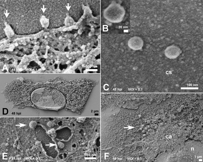
Ultrastructural analysis of SARS-CoV-2 interactions with the host cell via  high resolution scanning electron microscopy | Scientific Reports
Ultrastructural analysis of SARS-CoV-2 interactions with the host cell via high resolution scanning electron microscopy | Scientific Reports
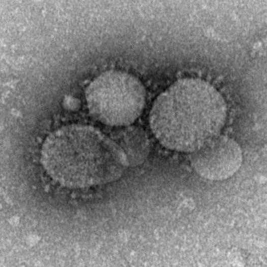
MERS-CoV Photos | CDC

Ultrastructural analysis of SARS-CoV-2 interactions with the host cell via  high resolution scanning electron microscopy | Scientific Reports
Ultrastructural analysis of SARS-CoV-2 interactions with the host cell via high resolution scanning electron microscopy | Scientific Reports

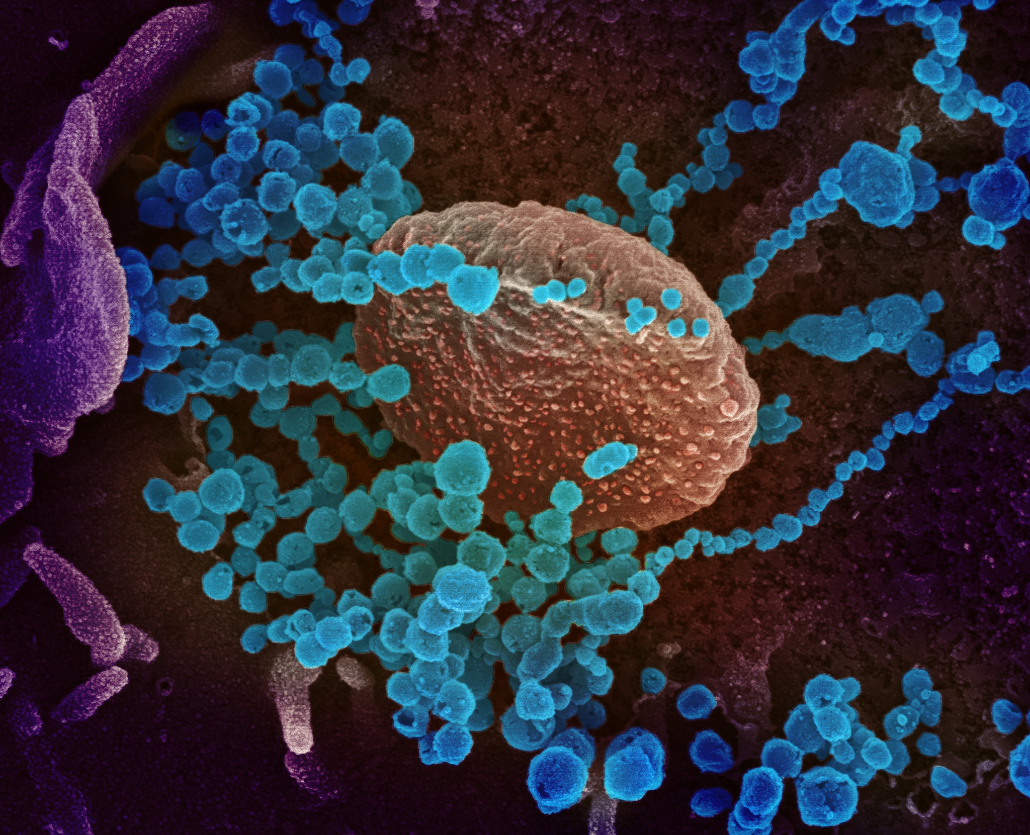
Novel coronavirus structure reveals targets for vaccines and treatments |  National Institutes of Health (NIH)
Novel coronavirus structure reveals targets for vaccines and treatments | National Institutes of Health (NIH)

Image Library | CDC Online Newsroom | CDC

Viruses | Special Issue : Electron Microscopy in Virus Diagnostics and  Research
Viruses | Special Issue : Electron Microscopy in Virus Diagnostics and Research

Electron microscopy of SARS-CoV-2: a challenging task - The Lancet

Viruses | Free Full-Text | Electron Microscopy in Discovery of Novel and  Emerging Viruses from the Collection of the World Reference Center for  Emerging Viruses and Arboviruses (WRCEVA) | HTML
Viruses | Free Full-Text | Electron Microscopy in Discovery of Novel and Emerging Viruses from the Collection of the World Reference Center for Emerging Viruses and Arboviruses (WRCEVA) | HTML

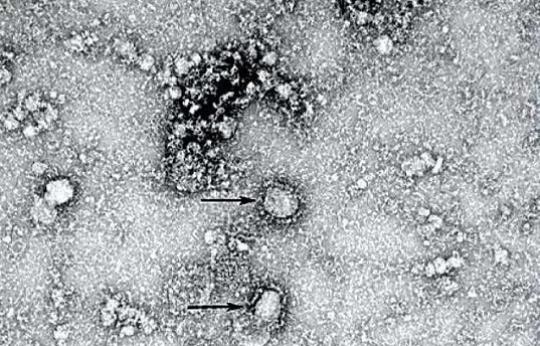
First electron microscopy images of new coronavirus - 2020 - Wiley  Analytical Science
First electron microscopy images of new coronavirus - 2020 - Wiley Analytical Science

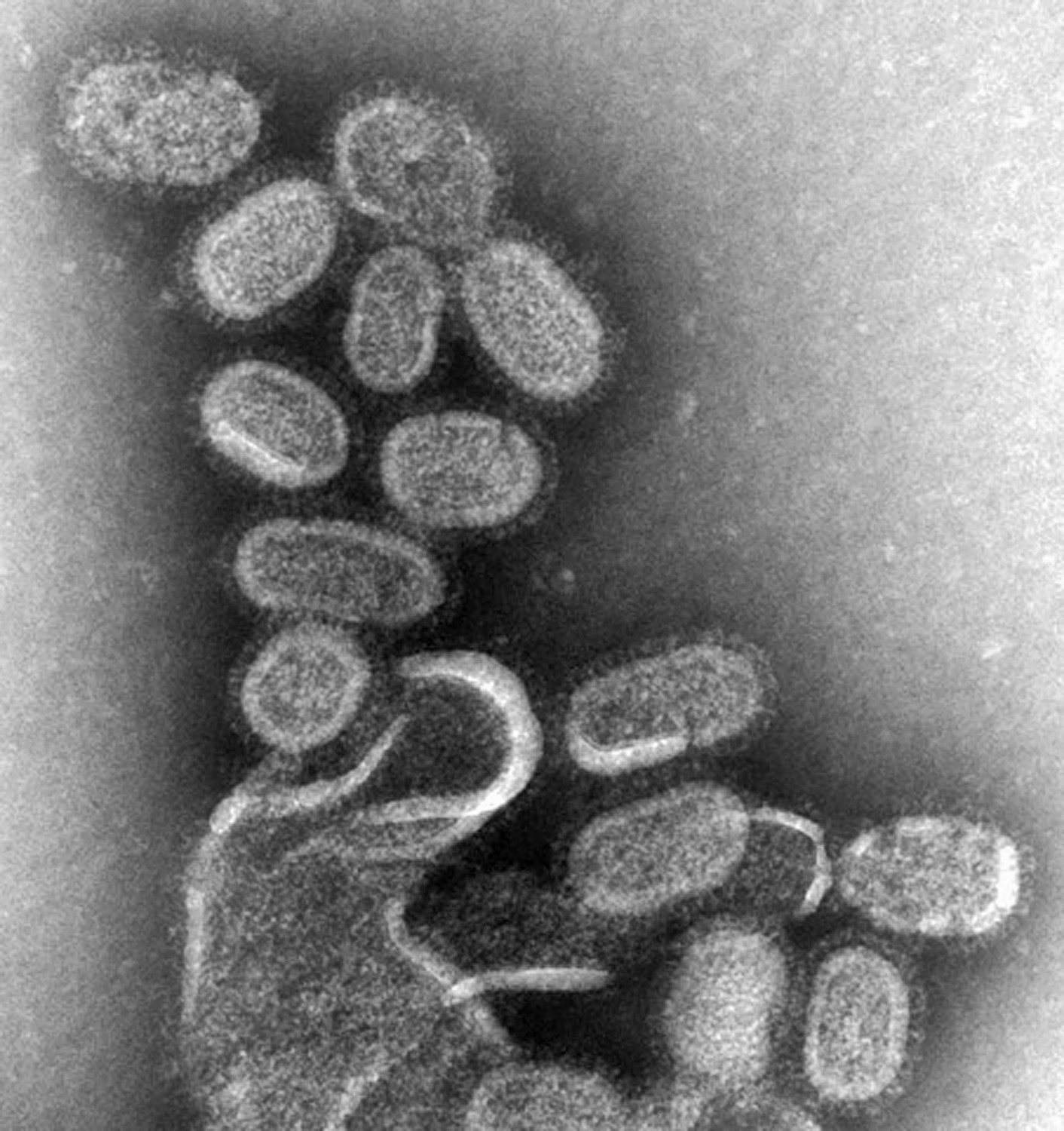
How To Catch The Flu (Under The Lens)

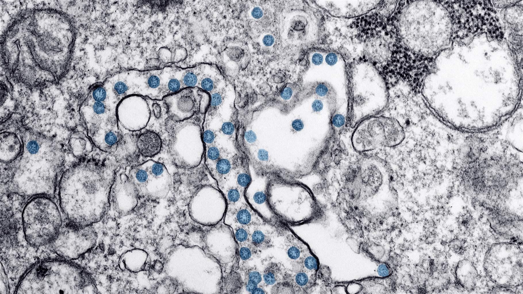
A Look at the Evolution of the Virus That Causes COVID-19 | The Pew  Charitable Trusts
A Look at the Evolution of the Virus That Causes COVID-19 | The Pew Charitable Trusts

CDC - Diagnosis: Electron Microscopy - Rabies

Electron Microscopy of Viruses - ScienceDirect

Correlative Scanning-Transmission Electron Microscopy Reveals that a  Chimeric Flavivirus Is Released as Individual Particles in Secretory  Vesicles | PLOS ONE
Correlative Scanning-Transmission Electron Microscopy Reveals that a Chimeric Flavivirus Is Released as Individual Particles in Secretory Vesicles | PLOS ONE
Structure that coronaviruses use to enter cells is unveiled | Newsroom

Electron microscopic investigations in COVID-19: not all crowns are coronas  - Kidney International
Electron microscopic investigations in COVID-19: not all crowns are coronas - Kidney International

Why misinterpretation of electron micrographs in SARS-CoV-2-infected tissue  goes viral - The Lancet
Why misinterpretation of electron micrographs in SARS-CoV-2-infected tissue goes viral - The Lancet

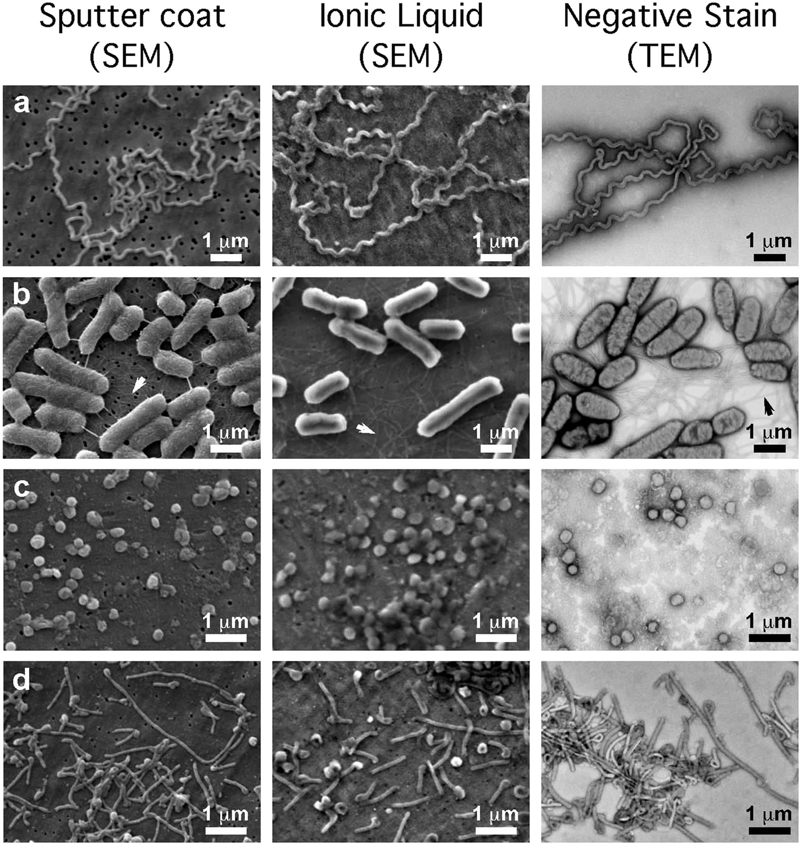
The scanning electron microscope in microbiology and diagnosis of  infectious disease | Scientific Reports
The scanning electron microscope in microbiology and diagnosis of infectious disease | Scientific Reports

How Oregon scientists found a new COVID-19 variant — and how we can stop it  - OPB
How Oregon scientists found a new COVID-19 variant — and how we can stop it - OPB

Multimedia Gallery - Transmission electron microscope image shows the virus  in spiny lobster blood cells. | NSF - National Science Foundation
Multimedia Gallery - Transmission electron microscope image shows the virus in spiny lobster blood cells. | NSF - National Science Foundation

Electron Cryo-Microscopy and Single-Particle Averaging of Rift Valley Fever  Virus: Evidence for GN-GC Glycoprotein Heterodimers | Journal of Virology
Electron Cryo-Microscopy and Single-Particle Averaging of Rift Valley Fever Virus: Evidence for GN-GC Glycoprotein Heterodimers | Journal of Virology

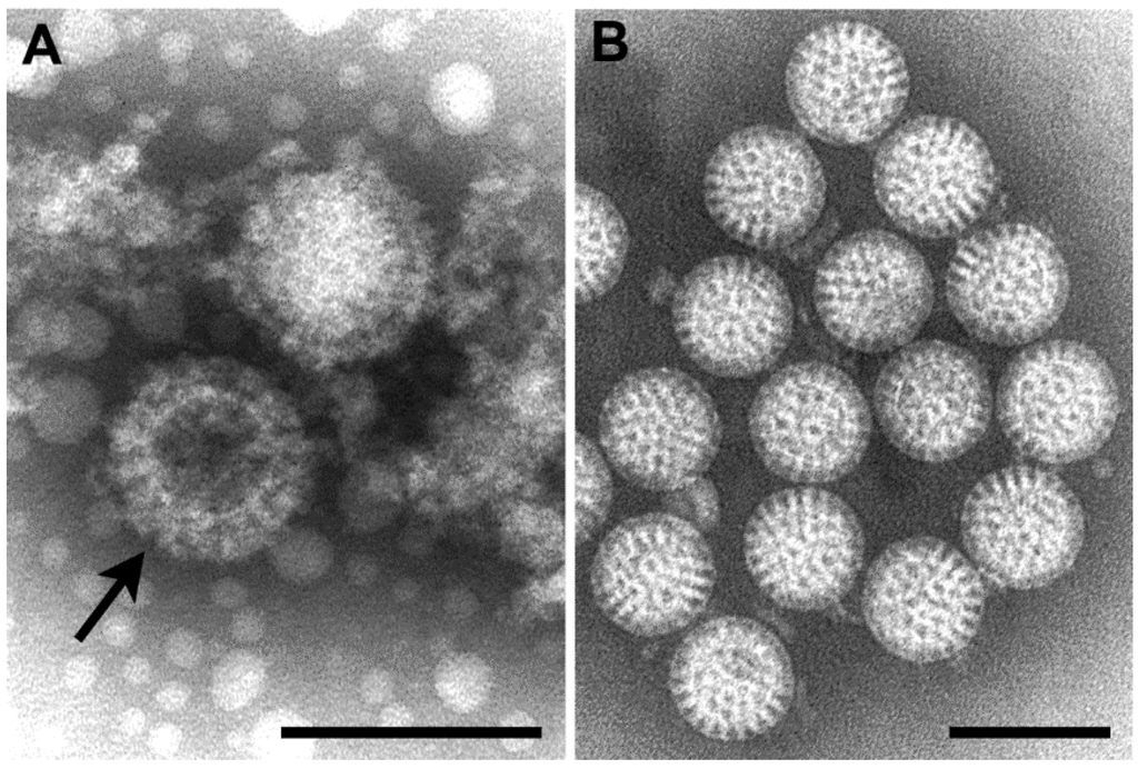
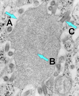
Identification of coronavirus particles by electron microscopy requires  demonstration of specific ultrastructural features | European Respiratory  Society
Identification of coronavirus particles by electron microscopy requires demonstration of specific ultrastructural features | European Respiratory Society
Chapter 25 Electron Microscopy of Viruses and Virus-Cell Interactions -  ScienceDirect
Chapter 25 Electron Microscopy of Viruses and Virus-Cell Interactions - ScienceDirect

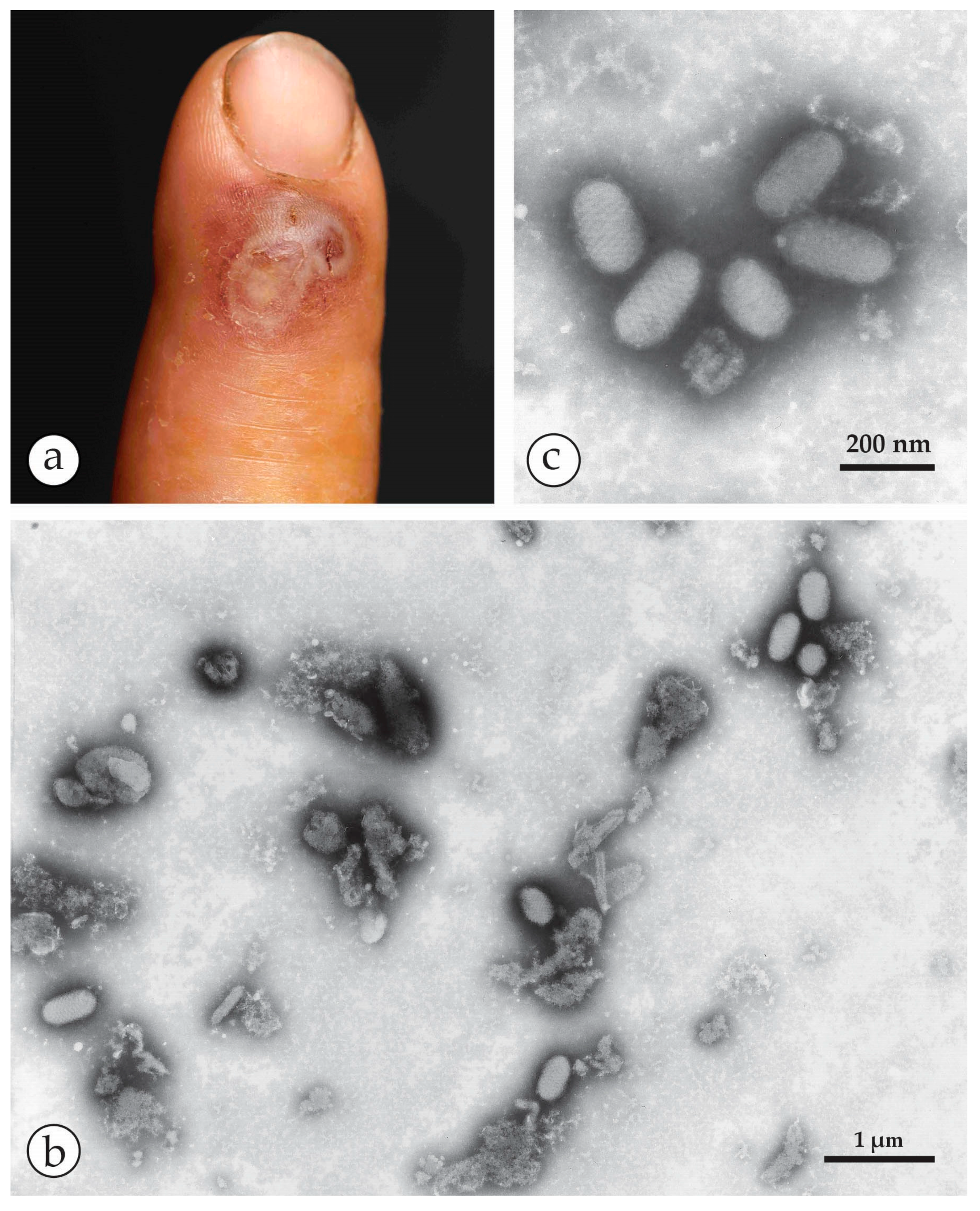
Viruses | Free Full-Text | Rapid Viral Diagnosis of Orthopoxviruses by  Electron Microscopy: Optional or a Must?
Viruses | Free Full-Text | Rapid Viral Diagnosis of Orthopoxviruses by Electron Microscopy: Optional or a Must?