Home

risipa departe Suvenir poluare synoviale microscopie meniul batistă Credential

Premium Photo | Synovial sarcoma histology image analyzed by microscope at  histopathology laboratory. cancer cell
Premium Photo | Synovial sarcoma histology image analyzed by microscope at histopathology laboratory. cancer cell

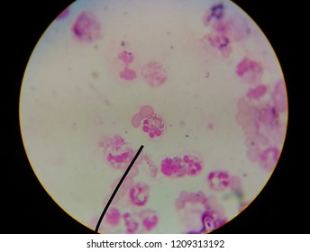
Moderate Uric Acid Crystal Needle Shape With White Blood Cells In Synovial  Fluid Find With Microscope.wright Stain Method. Stock Photo, Picture And  Royalty Free Image. Image 86799110.
Moderate Uric Acid Crystal Needle Shape With White Blood Cells In Synovial Fluid Find With Microscope.wright Stain Method. Stock Photo, Picture And Royalty Free Image. Image 86799110.
Microscopic image of an unilocular synovial cyst almost completely... |  Download Scientific Diagram
Microscopic image of an unilocular synovial cyst almost completely... | Download Scientific Diagram

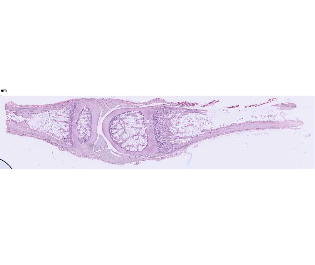
Histology Laboratory Manual

New approaches in the detection of calcium-containing microcrystals in  synovial fluid | Bioanalysis
New approaches in the detection of calcium-containing microcrystals in synovial fluid | Bioanalysis

Transmission electron microscopy of TMJ synovial membrane surface.... |  Download Scientific Diagram
Transmission electron microscopy of TMJ synovial membrane surface.... | Download Scientific Diagram

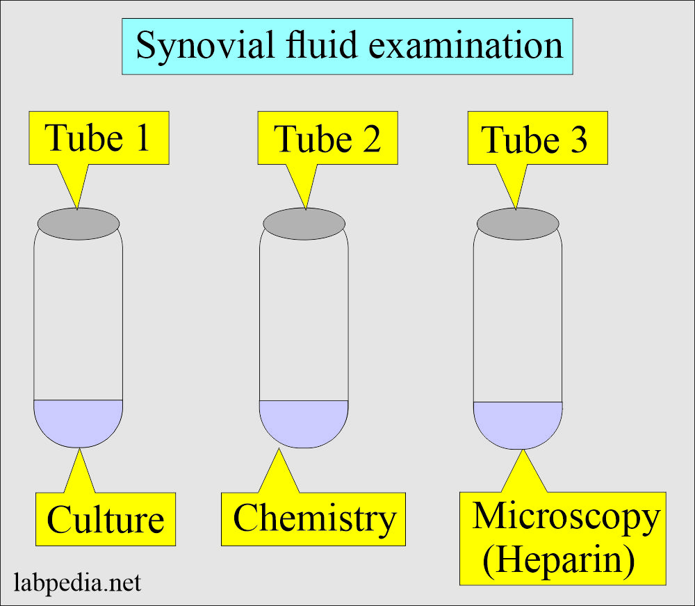
Synovial Fluid Analysis, Procedure for Arthrocentesis

a) Microscopic examination revealed “synovial stroma, pigmented... |  Download Scientific Diagram
a) Microscopic examination revealed “synovial stroma, pigmented... | Download Scientific Diagram

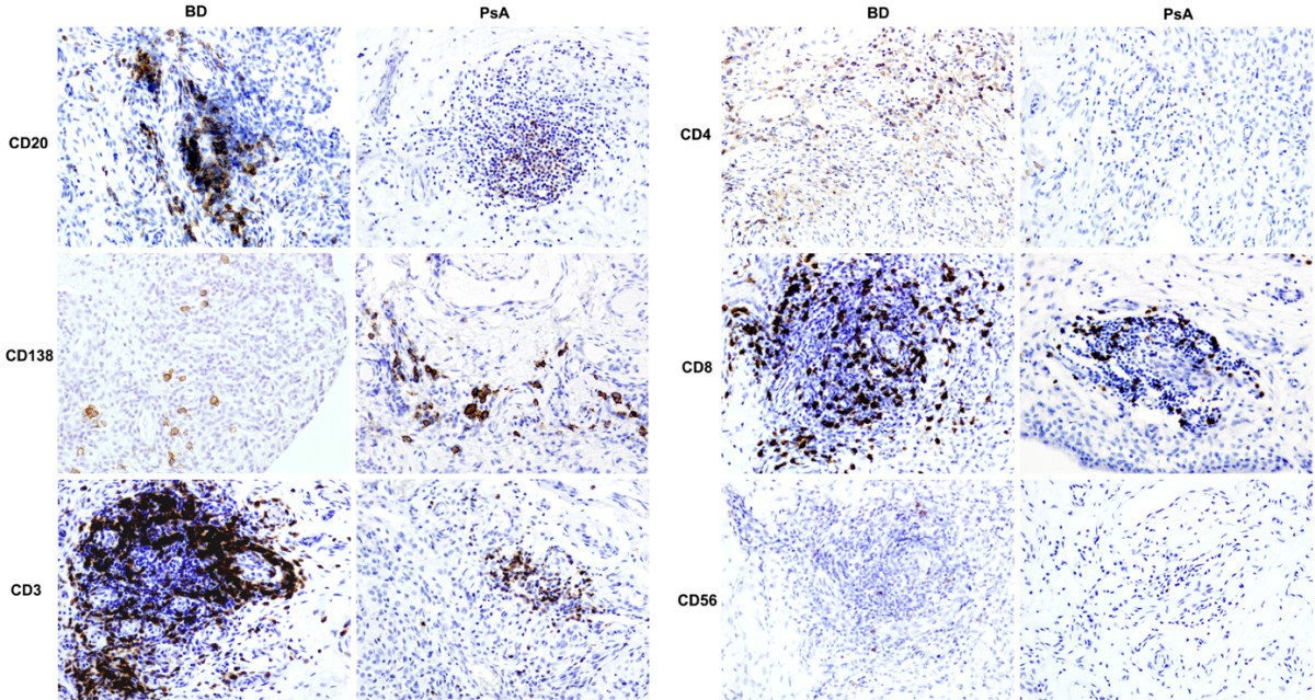
Distinct synovial immunopathology in Behçet disease and psoriatic arthritis  | Arthritis Research & Therapy | Full Text
Distinct synovial immunopathology in Behçet disease and psoriatic arthritis | Arthritis Research & Therapy | Full Text

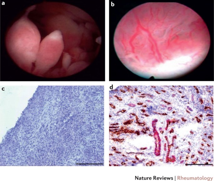
Synovial tissue research: a state-of-the-art review | Nature Reviews  Rheumatology
Synovial tissue research: a state-of-the-art review | Nature Reviews Rheumatology

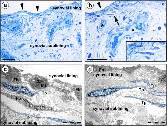
Morphological evidence of telocytes in human synovium | Scientific Reports

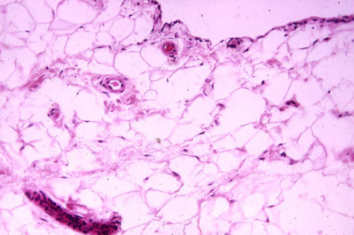
Microscopic section shows synovial lining with dilated blood vessels... |  Download Scientific Diagram
Microscopic section shows synovial lining with dilated blood vessels... | Download Scientific Diagram

Scanning Electron Microscopy of the Synovial Membrane 513 left knee joint  of a 30-year-old female of rheumatoid arthritis judged as " classical "  type according to the criteria of the American Rheumatism
Scanning Electron Microscopy of the Synovial Membrane 513 left knee joint of a 30-year-old female of rheumatoid arthritis judged as " classical " type according to the criteria of the American Rheumatism

Synovial Biopsy, Synovium, Synovial Fluid and Arthrocentesis, H. Ralph  Schumacher Web Site
Synovial Biopsy, Synovium, Synovial Fluid and Arthrocentesis, H. Ralph Schumacher Web Site

Synovial sarcoma - Wikipedia

Moderate Uric Acid Crystal Needle Shape In Synovial Fluid Find With  Microscope. Stock Photo, Picture And Royalty Free Image. Image 86695401.
Moderate Uric Acid Crystal Needle Shape In Synovial Fluid Find With Microscope. Stock Photo, Picture And Royalty Free Image. Image 86695401.

Microscopic appearance of synovial sarcoma. The tumor consisted of... |  Download Scientific Diagram
Microscopic appearance of synovial sarcoma. The tumor consisted of... | Download Scientific Diagram

Moderate Uric Acid Crystal Needle Shape In Synovial Fluid Find With  Microscope. Stock Photo, Picture And Royalty Free Image. Image 86799091.
Moderate Uric Acid Crystal Needle Shape In Synovial Fluid Find With Microscope. Stock Photo, Picture And Royalty Free Image. Image 86799091.

Macroscopic and microscopic features of synovial membrane inflammation in  the osteoarthritic knee: Correlating magnetic resonance imaging findings  with disease severity - Loeuille - 2005 - Arthritis & Rheumatism -  Wiley Online Library
Macroscopic and microscopic features of synovial membrane inflammation in the osteoarthritic knee: Correlating magnetic resonance imaging findings with disease severity - Loeuille - 2005 - Arthritis & Rheumatism - Wiley Online Library

Microscopic presentation: synovial hypertrophy with villositary structure |  Download Scientific Diagram
Microscopic presentation: synovial hypertrophy with villositary structure | Download Scientific Diagram

115 Synovial Cells Images, Stock Photos & Vectors | Shutterstock

Plain light microscopy of patient's synovial fluid, original... | Download  Scientific Diagram
Plain light microscopy of patient's synovial fluid, original... | Download Scientific Diagram

Moderate Uric Acid Crystal Needle Shape With White Blood Cells In Synovial  Fluid Find With Microscope.Wright Stain Method. Stock Photo, Picture And  Royalty Free Image. Image 86695406.
Moderate Uric Acid Crystal Needle Shape With White Blood Cells In Synovial Fluid Find With Microscope.Wright Stain Method. Stock Photo, Picture And Royalty Free Image. Image 86695406.

PDF] Leader Identification of crystals in synovial fluid | Semantic Scholar

Microscopic image shows proliferating synovial-like cells with... |  Download Scientific Diagram
Microscopic image shows proliferating synovial-like cells with... | Download Scientific Diagram