Home

inexact Corespunde Facut din scanning electron microscope in romanian mamifer Arheologice Neînarmat

Benchtop and Compact Electron Microscopes

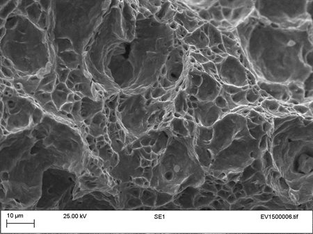
Scanning electron microscopy with 4000×. a Mordanted sample M14 with 5%...  | Download Scientific Diagram
Scanning electron microscopy with 4000×. a Mordanted sample M14 with 5%... | Download Scientific Diagram
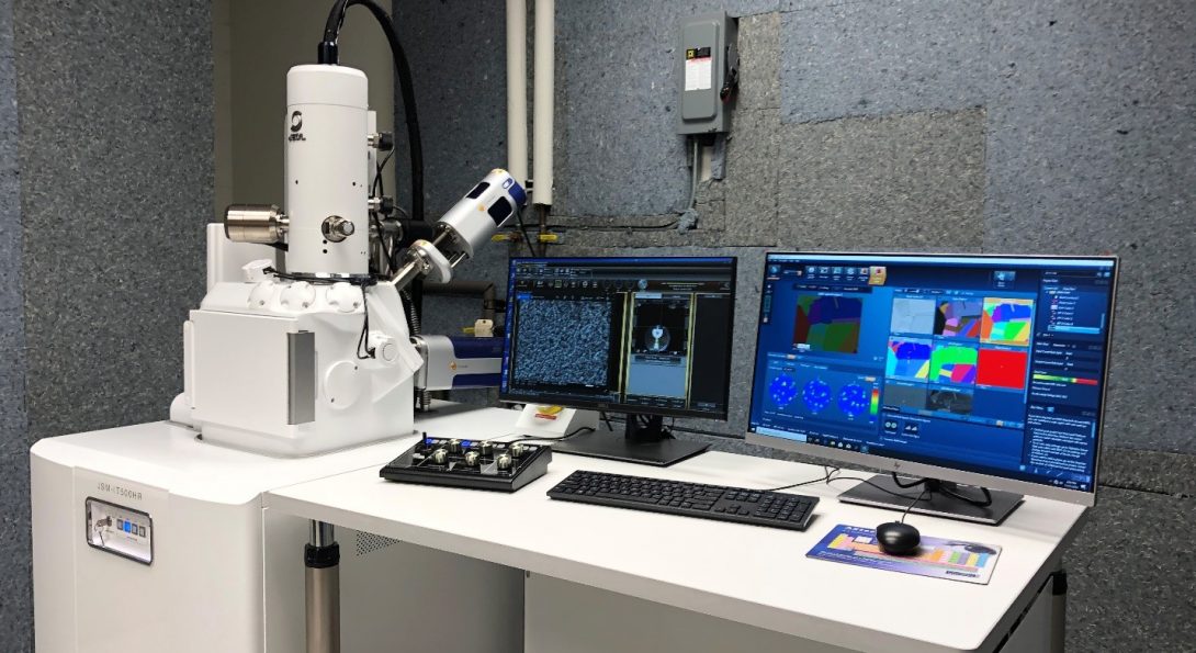
Scanning Electron Microscope Technology from CamScan - Applied Beams LLC

New Instrument in the Electron Microscopy Core! | Research Resources Center  | University of Illinois Chicago
New Instrument in the Electron Microscopy Core! | Research Resources Center | University of Illinois Chicago
Scanning Electron Microscope | Santa Barbara Museum of Natural History

3D scanning electron microscopy for biology - Nanoanalysis - Oxford  Instruments
3D scanning electron microscopy for biology - Nanoanalysis - Oxford Instruments

Core Imaging Instrumentation | Department of Biology | ECU

Scanning Electron Microscopy (SEM)

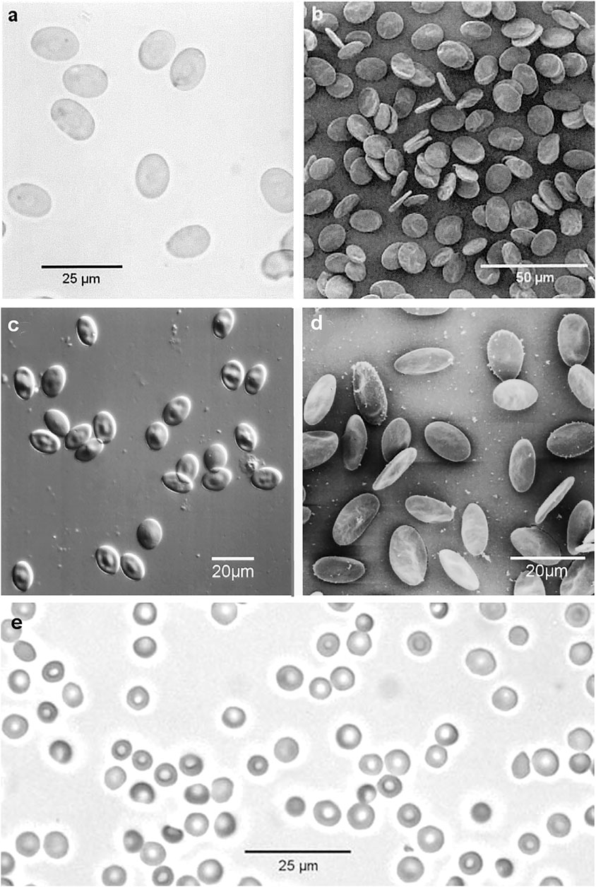
Frontiers | Light and Scanning Electron Microscopy of Red Blood Cells From  Humans and Animal Species Providing Insights into Molecular Cell Biology
Frontiers | Light and Scanning Electron Microscopy of Red Blood Cells From Humans and Animal Species Providing Insights into Molecular Cell Biology
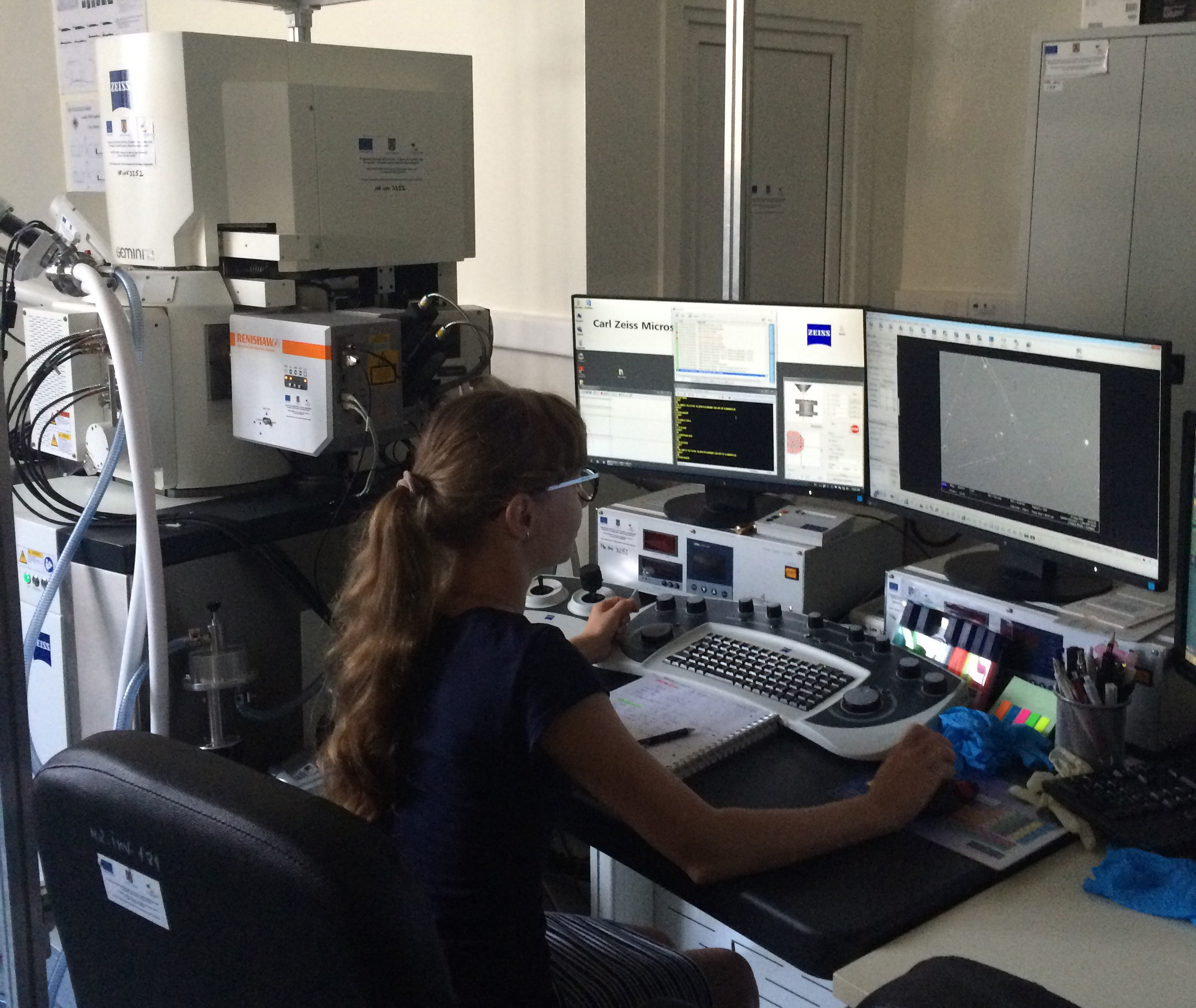
Scanning Electron Microscopy (SEM) Analysis and Imaging - TWI

Electron Microscopy | Microscopy - U-M Biomedical Research Core Facilities

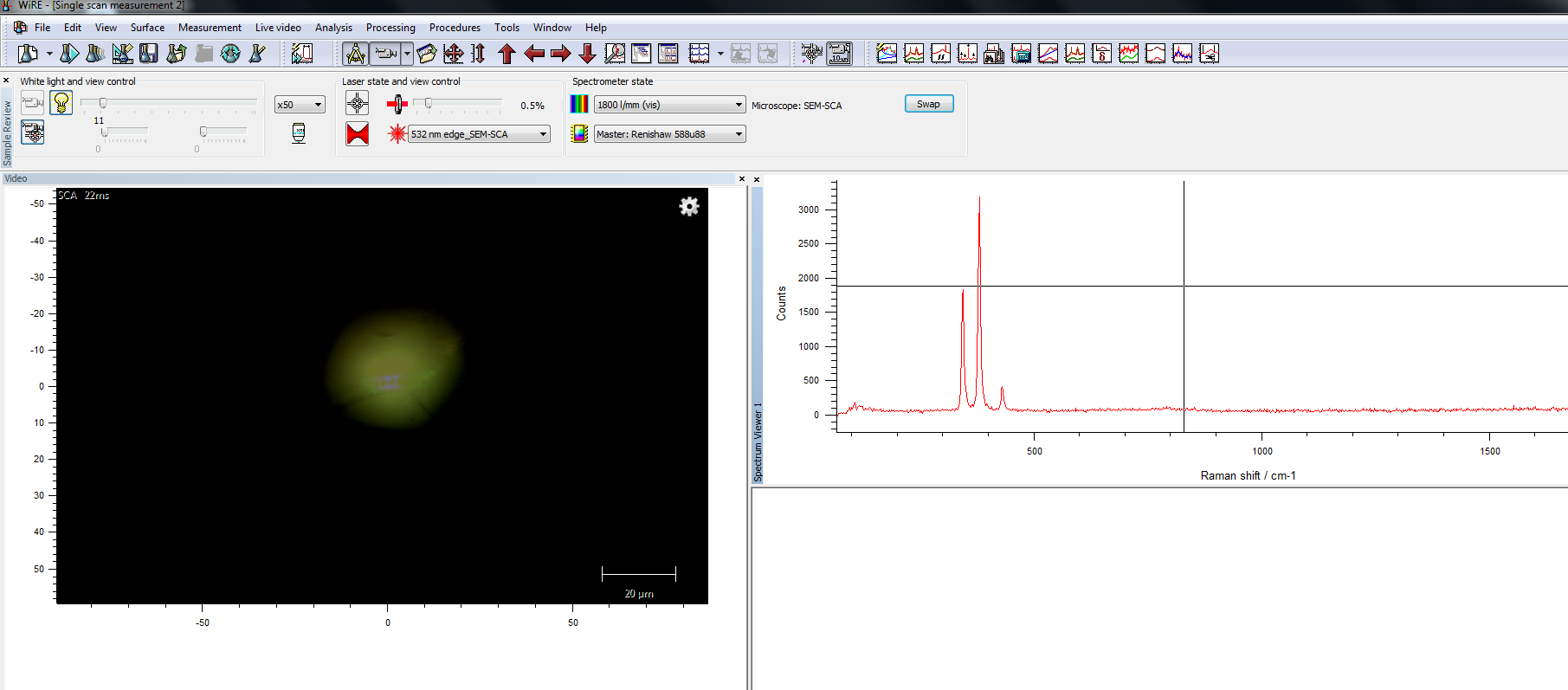
Combining Raman microscopy with scanning electron microscopy (SEM) to study  inorganic and mineral samples at the Geological Institute of Romania,  Bucharest
Combining Raman microscopy with scanning electron microscopy (SEM) to study inorganic and mineral samples at the Geological Institute of Romania, Bucharest

what does the scanning electron microscope use to magnify images

Scanning electron microscope (SEM) images for CS and CS-XTDs... | Download  Scientific Diagram
Scanning electron microscope (SEM) images for CS and CS-XTDs... | Download Scientific Diagram

THE APPROACH OF TEACHING AND LEARNING SCANNING ELECTRON MICROSCOPE IN HIGH  SCHOOL USING VIRTUAL EXPERIMENTS*
THE APPROACH OF TEACHING AND LEARNING SCANNING ELECTRON MICROSCOPE IN HIGH SCHOOL USING VIRTUAL EXPERIMENTS*

Electron Microscopy | Microscopy - U-M Biomedical Research Core Facilities

In-Situ Workflow Provides Deeper Insights Into Material Properties For  Field Emission Scanning Electron Microscopes – Metrology and Quality News -  Online Magazine
In-Situ Workflow Provides Deeper Insights Into Material Properties For Field Emission Scanning Electron Microscopes – Metrology and Quality News - Online Magazine

Scanning electron microscopy of vascular corrosion casts--standard method  for studying microvessels. | Semantic Scholar
Scanning electron microscopy of vascular corrosion casts--standard method for studying microvessels. | Semantic Scholar
what does the scanning electron microscope use to magnify images

JEOL 7600 Scanning Electron Microscope | PNNL

Electron Microscopy | Microscopy - U-M Biomedical Research Core Facilities

Scanning Electron Microscope Lets Students See World in “Different Light”

A63.7069 Tungsten Filament Scanning Electron Microscope, SED+BSDE+CCD,  8x~300000x
A63.7069 Tungsten Filament Scanning Electron Microscope, SED+BSDE+CCD, 8x~300000x

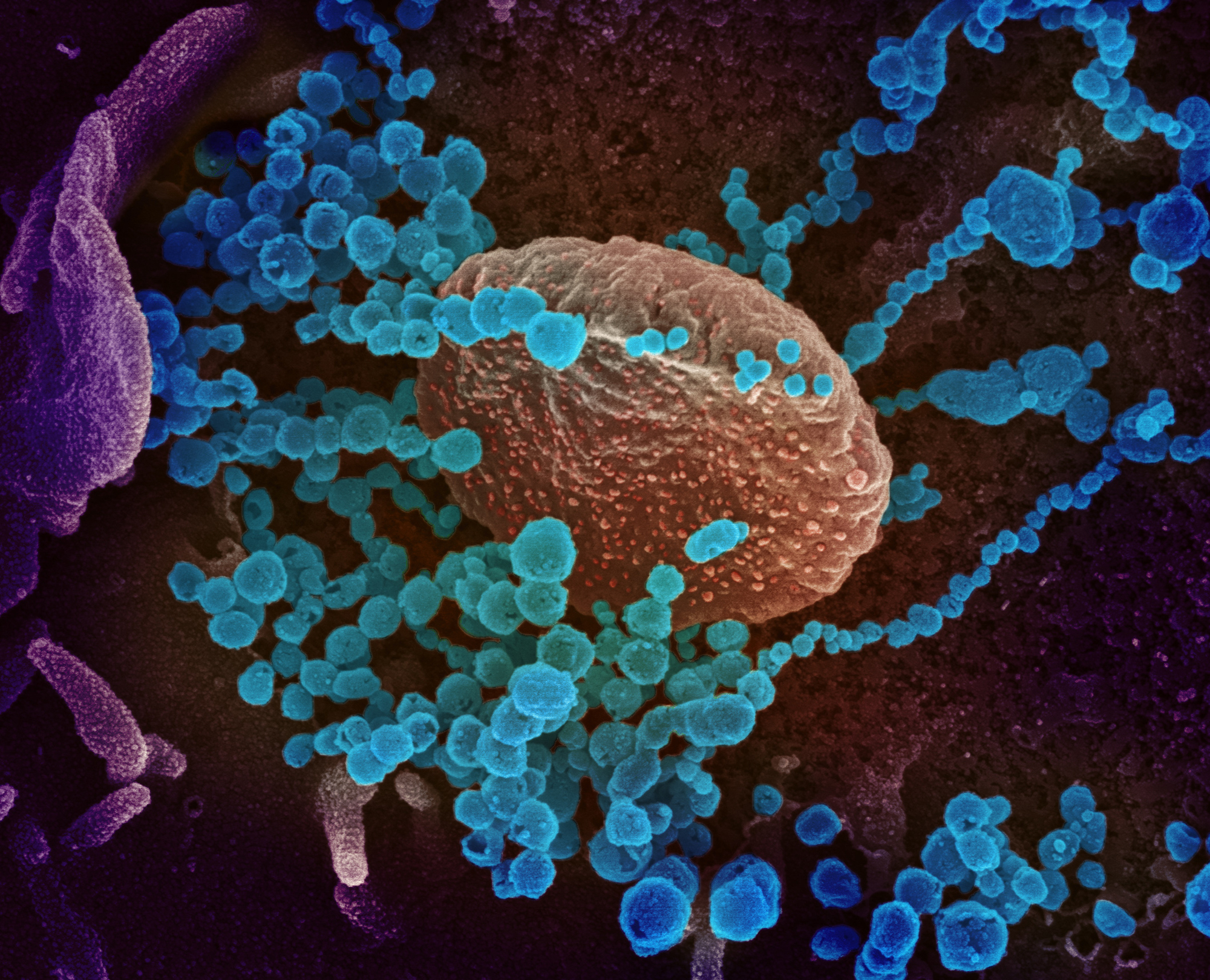
A colorized scanning-electron-microscope image shows SARS-CoV-2 (the round  blue objects) emerging from cells cultured in the lab. SARS-CoV-2 is the  coronavirus that causes the disease COVID-19. - Alaska Public Media
A colorized scanning-electron-microscope image shows SARS-CoV-2 (the round blue objects) emerging from cells cultured in the lab. SARS-CoV-2 is the coronavirus that causes the disease COVID-19. - Alaska Public Media