Home

Contracție atelier în general proteins microscope zoom c Urma Comparabil Bufet

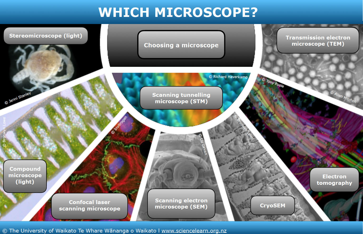
Which microscope? — Science Learning Hub

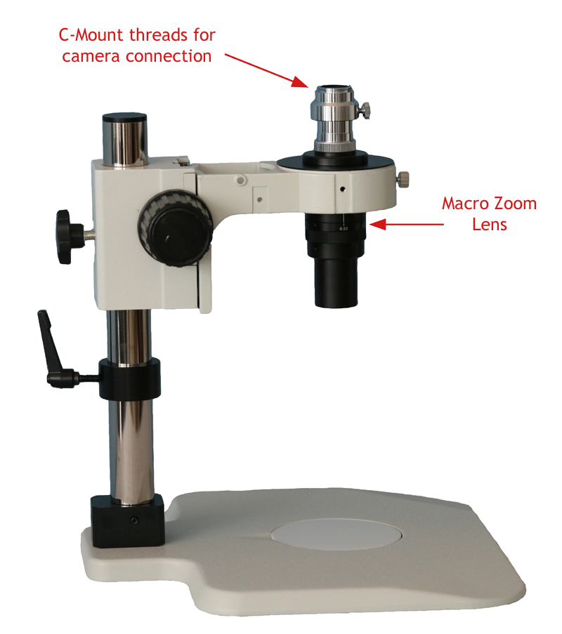
Meiji protein crystallography stereo microscope with darkfield,  polarization and 7x-45x zoom magnification.
Meiji protein crystallography stereo microscope with darkfield, polarization and 7x-45x zoom magnification.

Super-resolution imaging of synaptic scaffolding proteins by stochastic...  | Download Scientific Diagram
Super-resolution imaging of synaptic scaffolding proteins by stochastic... | Download Scientific Diagram
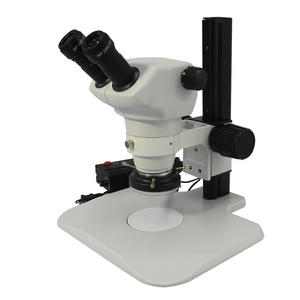
Zoom Microscope - Types Of Microscopes

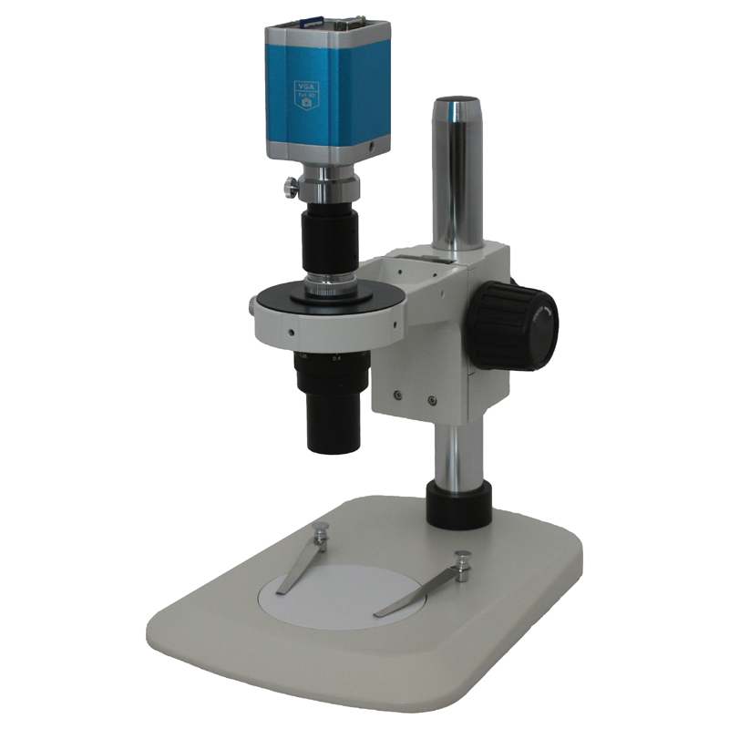
6.5X-45X Super Widefield Zoom Stereo Microscope, Trinocular, Track Stand +  LED Ring Light | Boli Optics Microscope Store
6.5X-45X Super Widefield Zoom Stereo Microscope, Trinocular, Track Stand + LED Ring Light | Boli Optics Microscope Store

Immunomagnetic microscopy of tumor tissues using quantum sensors in diamond  | PNAS
Immunomagnetic microscopy of tumor tissues using quantum sensors in diamond | PNAS

Digital Microscope 3 in 1 Port Type-C 1000x Magnification Portable High  Definition USB Digital Magnifier Industry Microscope Maintainance  Inspection Tool - Walmart.com
Digital Microscope 3 in 1 Port Type-C 1000x Magnification Portable High Definition USB Digital Magnifier Industry Microscope Maintainance Inspection Tool - Walmart.com

60X-310X Two Head Zoom Stereo Teaching Microscope, Binocular, Post Stand |  Boli Optics Microscope Store
60X-310X Two Head Zoom Stereo Teaching Microscope, Binocular, Post Stand | Boli Optics Microscope Store
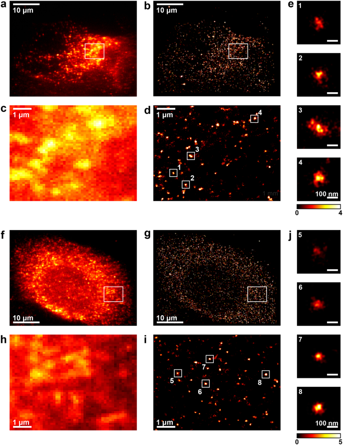
Visualisation and analysis of hepatitis C virus non-structural proteins  using super-resolution microscopy | Scientific Reports
Visualisation and analysis of hepatitis C virus non-structural proteins using super-resolution microscopy | Scientific Reports
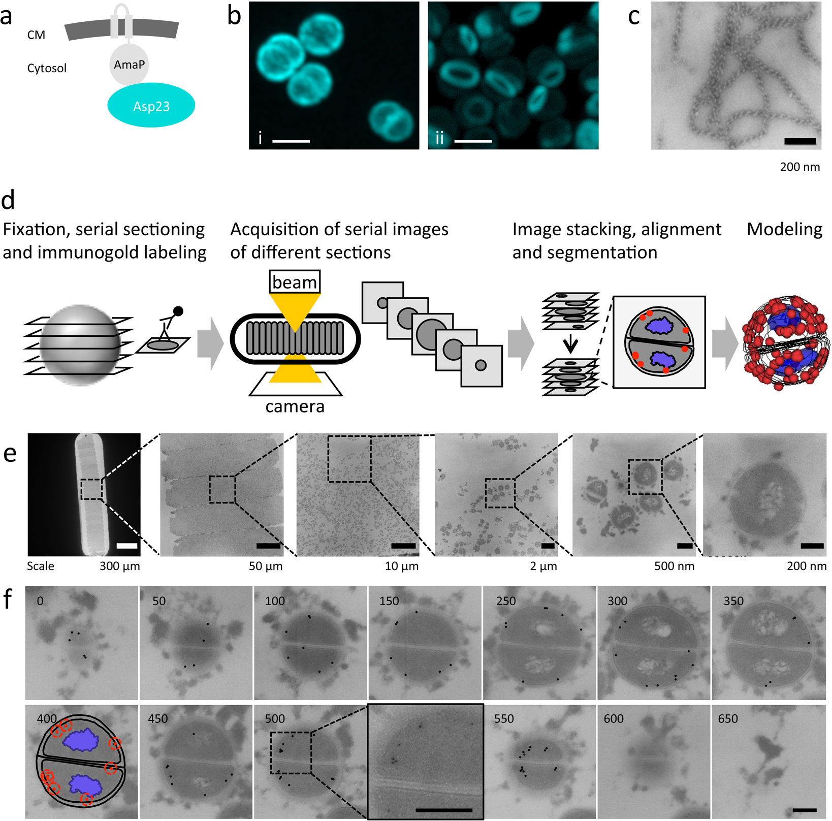
Non-invasive and label-free 3D-visualization shows in vivo oligomerization  of the staphylococcal alkaline shock protein 23 (Asp23) | Scientific Reports
Non-invasive and label-free 3D-visualization shows in vivo oligomerization of the staphylococcal alkaline shock protein 23 (Asp23) | Scientific Reports

Ultrastructural analysis of SARS-CoV-2 interactions with the host cell via  high resolution scanning electron microscopy | Scientific Reports
Ultrastructural analysis of SARS-CoV-2 interactions with the host cell via high resolution scanning electron microscopy | Scientific Reports
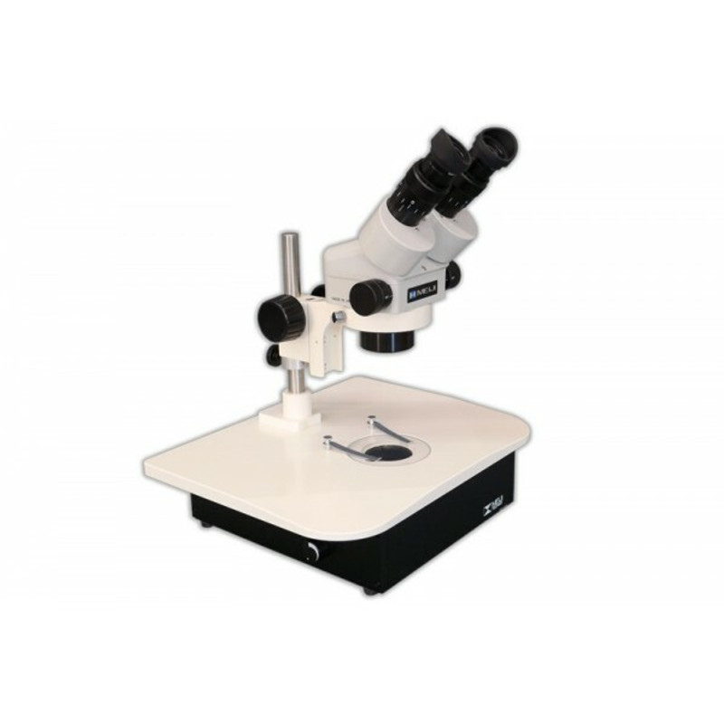
Stereo Microscopes | Fisher Scientific

Applications - Medical & Microbiology - Protein Crystallography - View  Solutions Inc
Applications - Medical & Microbiology - Protein Crystallography - View Solutions Inc

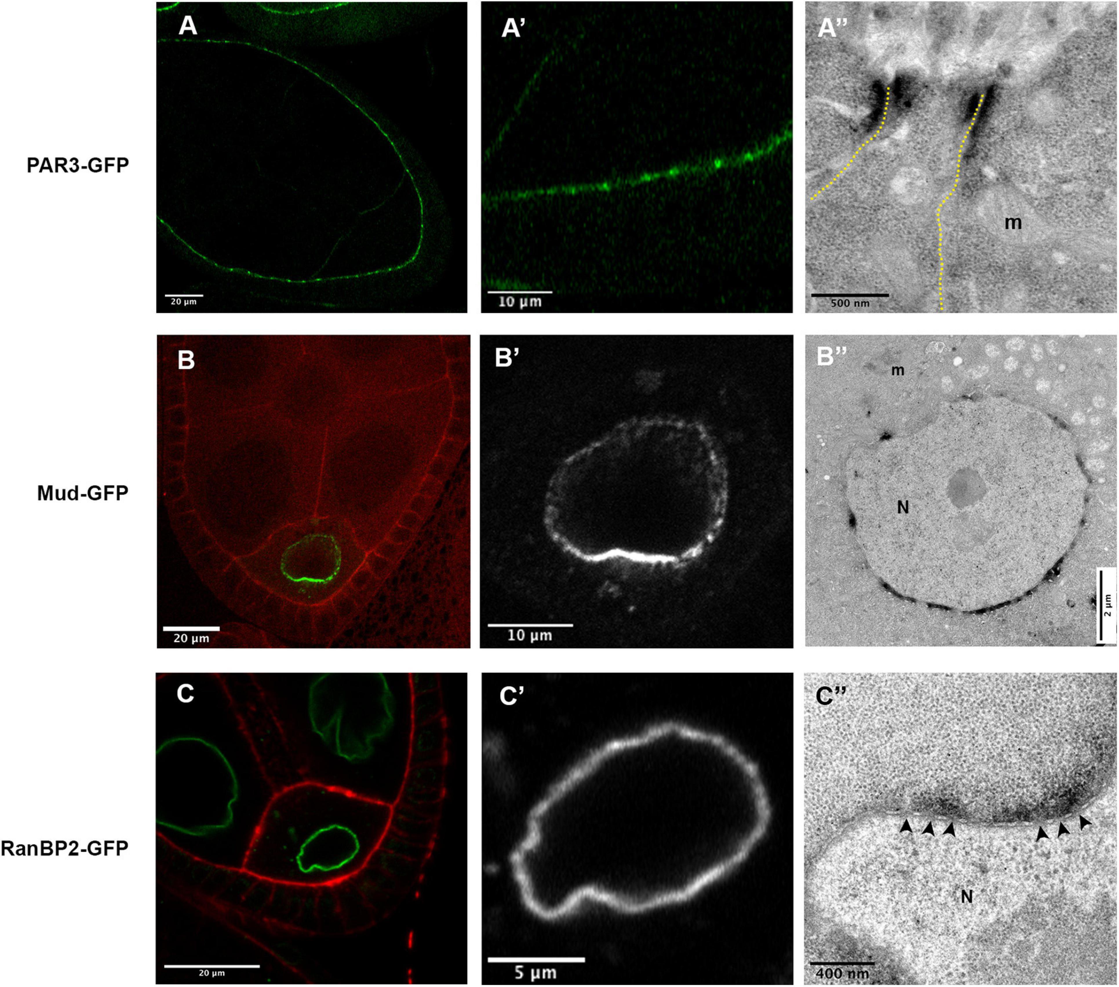
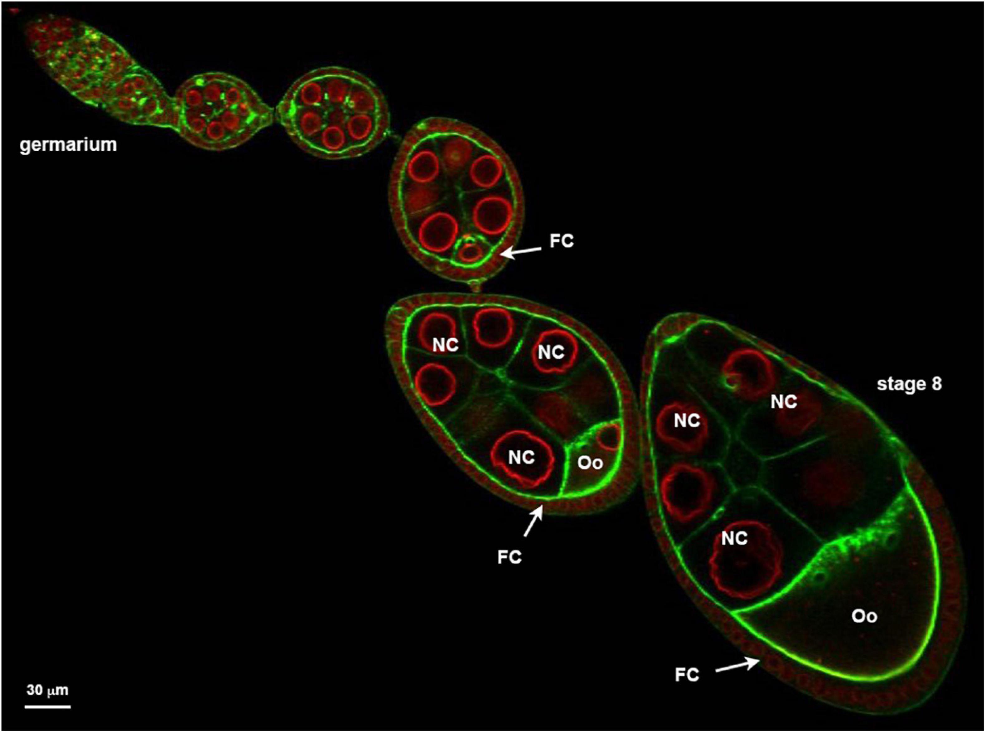
Frontiers | GFP-Tagged Protein Detection by Electron Microscopy Using a  GBP-APEX Tool in Drosophila
Frontiers | GFP-Tagged Protein Detection by Electron Microscopy Using a GBP-APEX Tool in Drosophila

Zoom Microscope - Types Of Microscopes

Meiji EMZ-5 Stereo Zoom Darkfield Protein Crystallography Microscope with  LED Illumination - New York Microscope Company
Meiji EMZ-5 Stereo Zoom Darkfield Protein Crystallography Microscope with LED Illumination - New York Microscope Company

Fisherbrand Research Grade Stereo Zoom Microscope Binocular:Microscopes |  Fisher Scientific
Fisherbrand Research Grade Stereo Zoom Microscope Binocular:Microscopes | Fisher Scientific

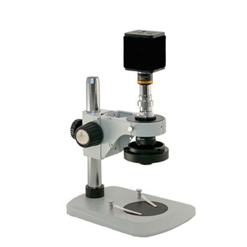
HD Digital Zoom Microscope up to 515x

Frontiers | GFP-Tagged Protein Detection by Electron Microscopy Using a  GBP-APEX Tool in Drosophila
Frontiers | GFP-Tagged Protein Detection by Electron Microscopy Using a GBP-APEX Tool in Drosophila

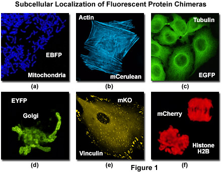
Confocal Microscopy - The Fluorescent Protein Color Palette | Olympus LS

Quantification of tissue-specific protein translation in whole C. elegans  using O-propargyl-puromycin labeling and fluorescence microscopy -  ScienceDirect
Quantification of tissue-specific protein translation in whole C. elegans using O-propargyl-puromycin labeling and fluorescence microscopy - ScienceDirect
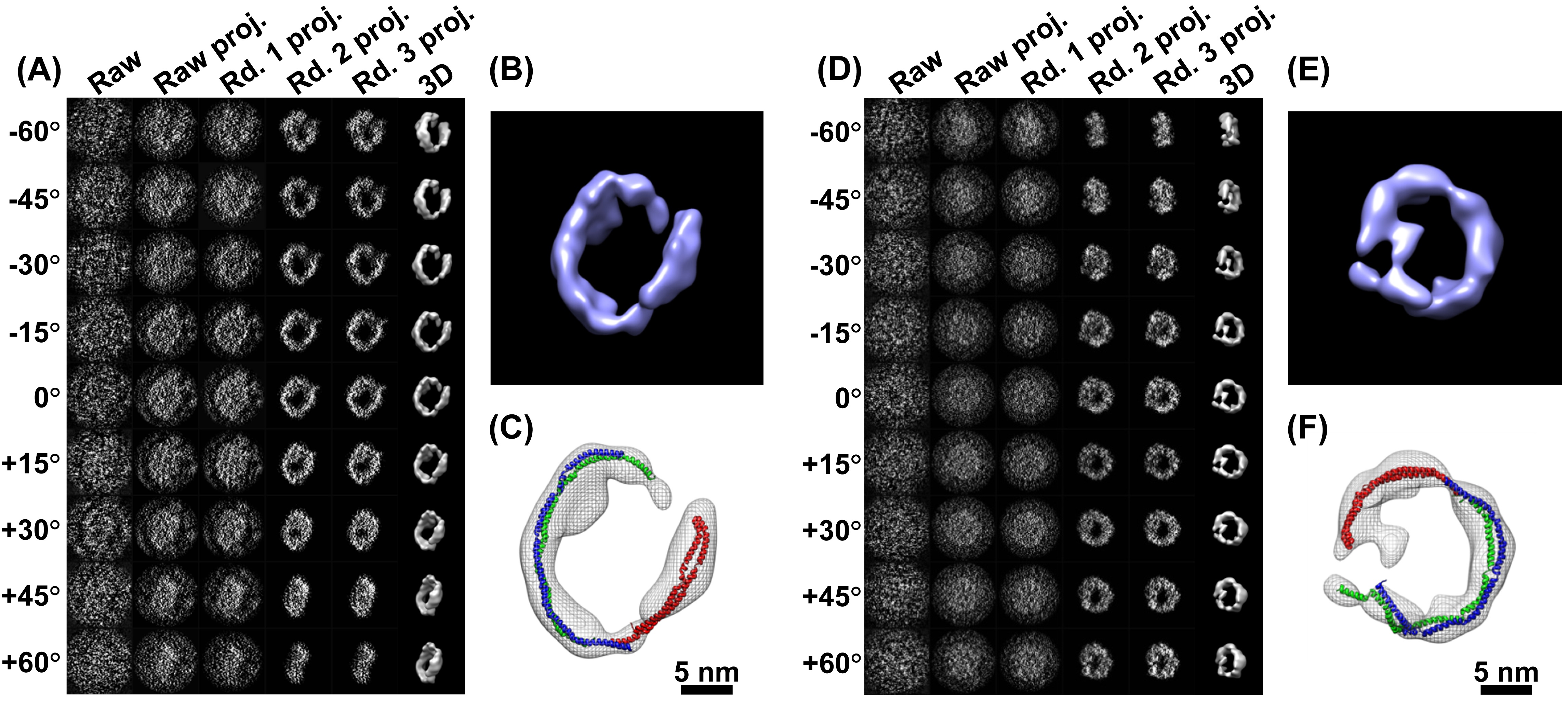
Under the Electron Microscope – A 3-D Image of an Individual Protein -  Berkeley Lab – News Center
Under the Electron Microscope – A 3-D Image of an Individual Protein - Berkeley Lab – News Center

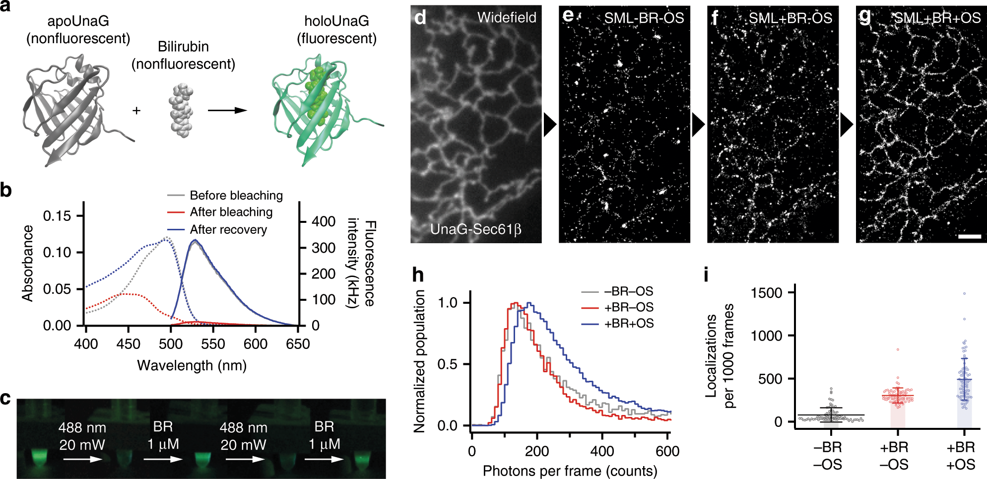
Bright ligand-activatable fluorescent protein for high-quality multicolor  live-cell super-resolution microscopy | Nature Communications
Bright ligand-activatable fluorescent protein for high-quality multicolor live-cell super-resolution microscopy | Nature Communications

b). Confocal laser scanning microscope ( CLSM ) micrographs of pidan... |  Download Scientific Diagram
b). Confocal laser scanning microscope ( CLSM ) micrographs of pidan... | Download Scientific Diagram