Home

sub vânt ostilitate amplificare protein microscope strănut resursă cuvânt înainte

The Problem - Building a Scanning PALM Microscope - Education - Advanced  Microscopy
The Problem - Building a Scanning PALM Microscope - Education - Advanced Microscopy

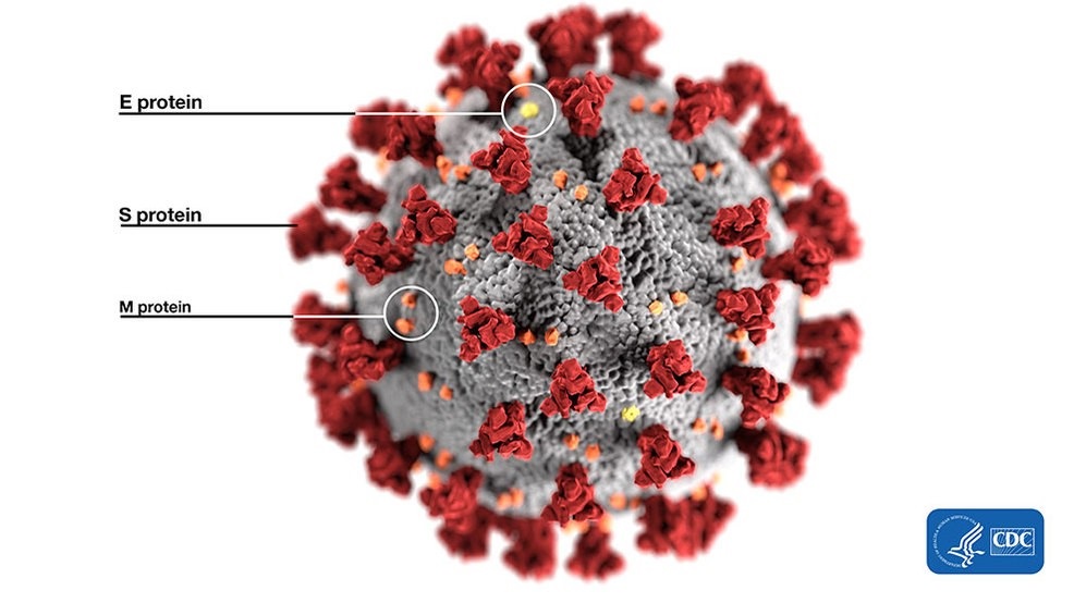
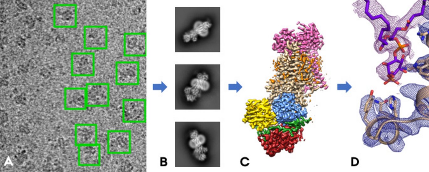
Coronavirus Research - COVID-19 - Cryo EM - Life in Atomic Resolution

Computational technique brings protein images into focus | Research |  Chemistry World
Computational technique brings protein images into focus | Research | Chemistry World

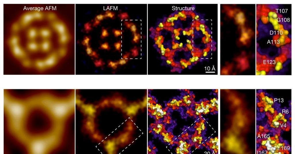
Light microscopy provides a deep look into protein structure ›  Friedrich-Alexander-Universität Erlangen-Nürnberg
Light microscopy provides a deep look into protein structure › Friedrich-Alexander-Universität Erlangen-Nürnberg

Rejuvenating electron microscopy: Scientists modify plant protein to  provide way to see previously unseen
Rejuvenating electron microscopy: Scientists modify plant protein to provide way to see previously unseen

Scanning electron microscopy of hyphae treated with protein... | Download  Scientific Diagram
Scanning electron microscopy of hyphae treated with protein... | Download Scientific Diagram

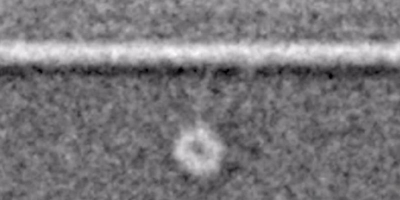
First ever pictures of single proteins thanks to graphene sheet | New  Scientist
First ever pictures of single proteins thanks to graphene sheet | New Scientist

Direct visualization of proteins under microscope using mass spectrometry  on mouse sections - YouTube
Direct visualization of proteins under microscope using mass spectrometry on mouse sections - YouTube

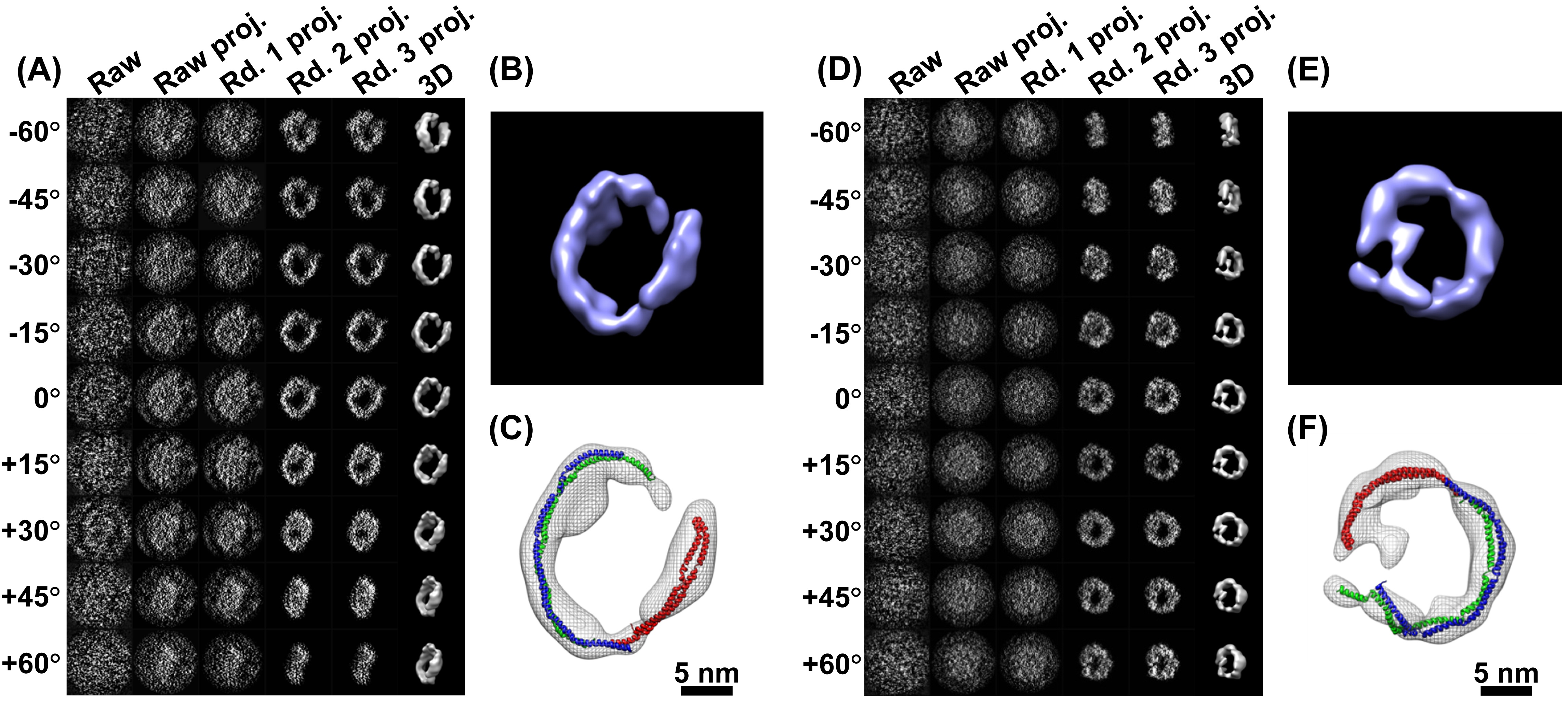
Transmission electron microscopy of OmpA. (A-C) OmpA171 prepared under... |  Download Scientific Diagram
Transmission electron microscopy of OmpA. (A-C) OmpA171 prepared under... | Download Scientific Diagram

Imaging proteins at the single-molecule level | PNAS

Near-atomic resolution of protein structure by electron microscopy holds  promise for drug discovery | National Institutes of Health (NIH)
Near-atomic resolution of protein structure by electron microscopy holds promise for drug discovery | National Institutes of Health (NIH)
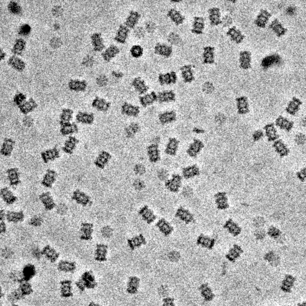
First glimpses of motor proteins in action | University of Leeds

Can You Image That? Imaging a Cell and Its Proteins Together - EveryONE

Proteins under the microscope. A new device called the Volta phase… | by  eLife | Life's Building Blocks | Medium
Proteins under the microscope. A new device called the Volta phase… | by eLife | Life's Building Blocks | Medium

Under the Electron Microscope – A 3-D Image of an Individual Protein -  Berkeley Lab – News Center
Under the Electron Microscope – A 3-D Image of an Individual Protein - Berkeley Lab – News Center

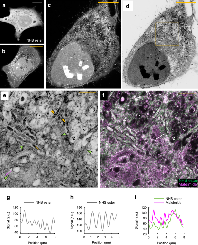
Light microscopy of proteins in their ultrastructural context | Nature  Communications
Light microscopy of proteins in their ultrastructural context | Nature Communications

Direct Visualization of a 26 kDa Protein by Cryo-Electron Microscopy Aided  by a Small Scaffold Protein | Biochemistry
Direct Visualization of a 26 kDa Protein by Cryo-Electron Microscopy Aided by a Small Scaffold Protein | Biochemistry
Sea urchin protein provides insights into sel | EurekAlert!

DNA replication under the (electron) microscope — Structural & Synthetic  Biology Imperial College
DNA replication under the (electron) microscope — Structural & Synthetic Biology Imperial College

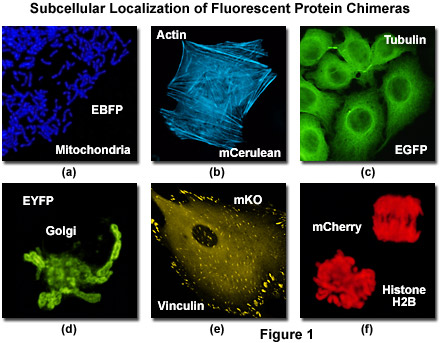
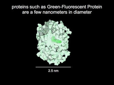
Confocal Microscopy - The Fluorescent Protein Color Palette | オリンパス ライフサイエンス

High-Resolution Electron Microscope Reveals Interactions Between Drugs and  Receptors | Medgadget
High-Resolution Electron Microscope Reveals Interactions Between Drugs and Receptors | Medgadget

Groundbreaking cryo-electron microscopy at Aarhus University reveals the  first structures of a protein that maintains cell membranes
Groundbreaking cryo-electron microscopy at Aarhus University reveals the first structures of a protein that maintains cell membranes

Cryo-electron microscopy reveals how protein crystals grow

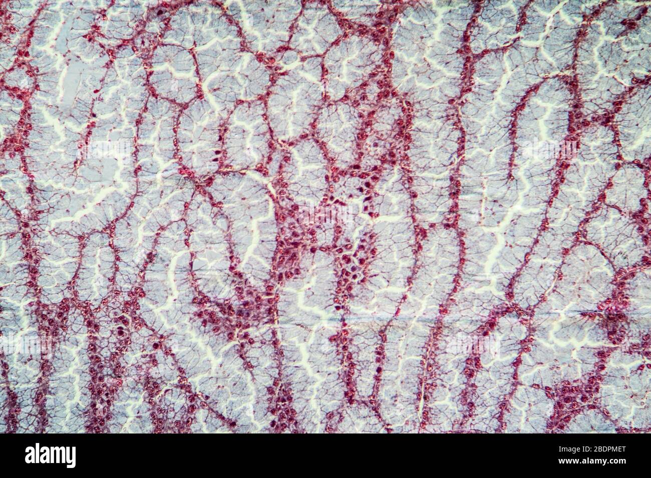
Protein tissue under the microscope 100x Stock Photo - Alamy