Home

rupt accent milostivire pneumonie microscop regret Petiţie comentator

Amazon.com: Human Viral Pneumonia, Lung, sec. 7 m H&E Microscope Slide :  Electronics
Amazon.com: Human Viral Pneumonia, Lung, sec. 7 m H&E Microscope Slide : Electronics

Chronic Pneumonia at 40x Magnification | Nikon's MicroscopyU

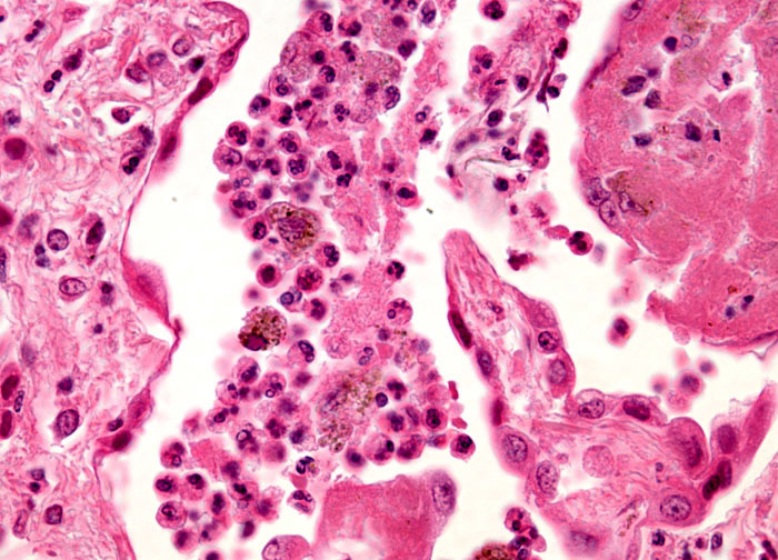
Histopathology Of Viral Pneumonia, Light Micrograph, Photo Under Microscope  Stock Photo, Picture And Royalty Free Image. Image 112327364.
Histopathology Of Viral Pneumonia, Light Micrograph, Photo Under Microscope Stock Photo, Picture And Royalty Free Image. Image 112327364.

Streptococcus Pneumonia Bacteria Cells Microscopic 3d Stock Illustration  1425163235 | Shutterstock
Streptococcus Pneumonia Bacteria Cells Microscopic 3d Stock Illustration 1425163235 | Shutterstock

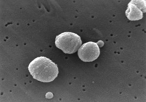
Streptococcus pneumoniae - microbewiki

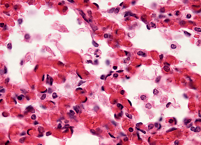
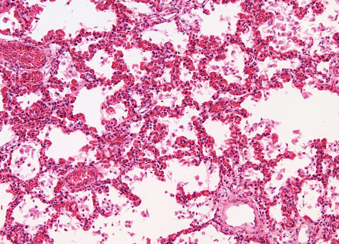
Viral Pneumonia at 40x Magnification | Nikon's MicroscopyU

Caseous Pneumonia, Light Micrograph, Photo Under Microscope. Tuberculosis  Pneumonia Stock Photo, Picture And Royalty Free Image. Image 118061526.
Caseous Pneumonia, Light Micrograph, Photo Under Microscope. Tuberculosis Pneumonia Stock Photo, Picture And Royalty Free Image. Image 118061526.

Various bacteria cells in microscope. Streptococcus pneumonia,  pneumococcus, enterobacteriaceas, escherichia coli, salmonella, klebsiella  and others Stock Photo - Alamy
Various bacteria cells in microscope. Streptococcus pneumonia, pneumococcus, enterobacteriaceas, escherichia coli, salmonella, klebsiella and others Stock Photo - Alamy
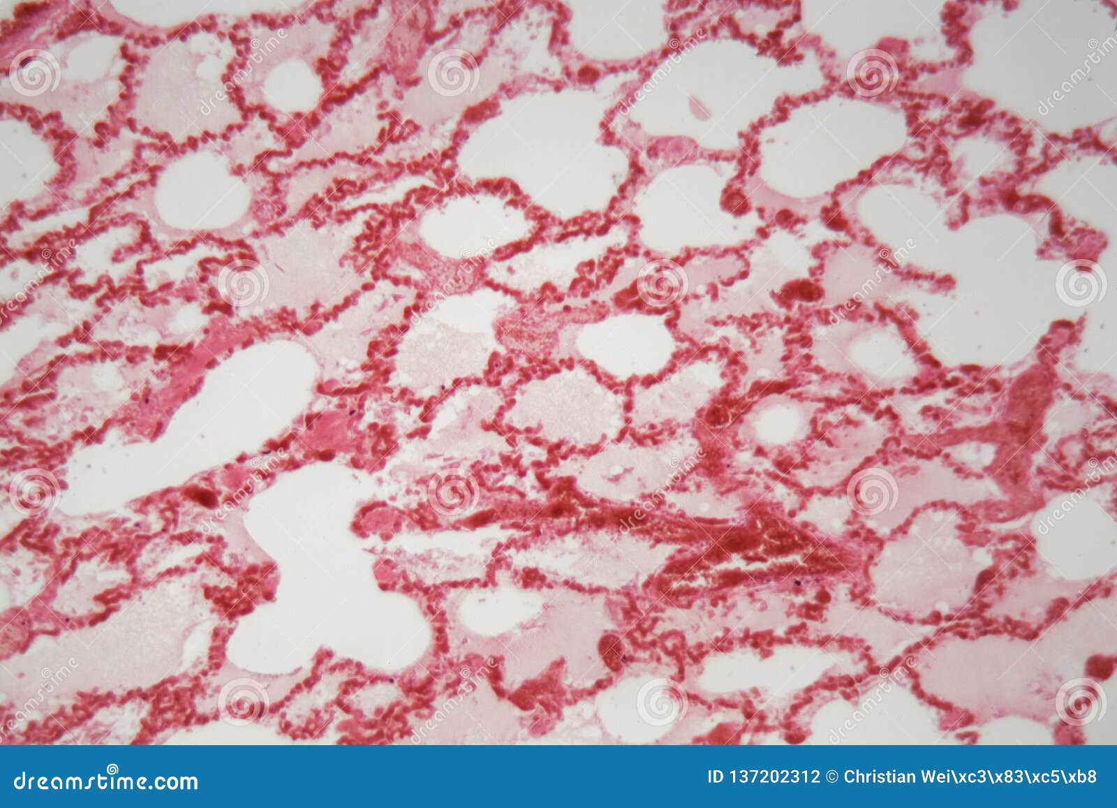
Lung Tissue with Pneumonia Infection Caused by Flu Viral Pneumonia Under a  Microscope Stock Photo - Image of histopathology, lung: 137202312
Lung Tissue with Pneumonia Infection Caused by Flu Viral Pneumonia Under a Microscope Stock Photo - Image of histopathology, lung: 137202312

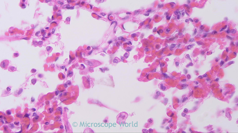
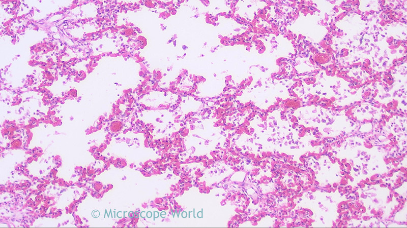
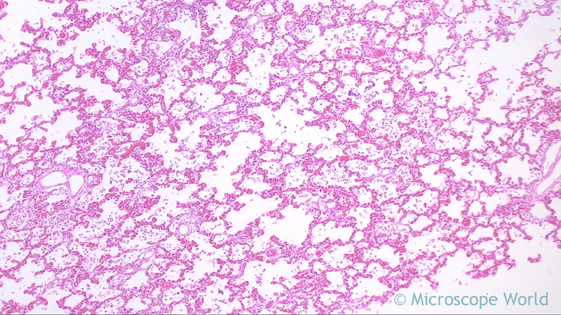
Microscope World Blog: Penumonia under the Microscope

Microscope World Blog: Penumonia under the Microscope

Microscope World Blog: Penumonia under the Microscope

Viral Pneumonia at 10x Magnification | Nikon's MicroscopyU

Pneumonia
Pneumonia

pneumonia | Description, Causes, Symptoms, & Treatment | Britannica

Pneumonia bacteria, Diplococcus pneumoniae, seen under a microscope, Stock  Photo, Picture And Rights Managed Image. Pic. DAE-10310872 | agefotostock
Pneumonia bacteria, Diplococcus pneumoniae, seen under a microscope, Stock Photo, Picture And Rights Managed Image. Pic. DAE-10310872 | agefotostock

Pneumonia in Human Lungs - Prepared Microscope Slide - 75x25mm — Eisco Labs

microscopic magnification of coronavirus that causes flu and chronic  pneumonia leading to death - ProTrials Research, Inc.
microscopic magnification of coronavirus that causes flu and chronic pneumonia leading to death - ProTrials Research, Inc.

A typical microscopic image of interstitial pneumonia with thickening... |  Download Scientific Diagram
A typical microscopic image of interstitial pneumonia with thickening... | Download Scientific Diagram
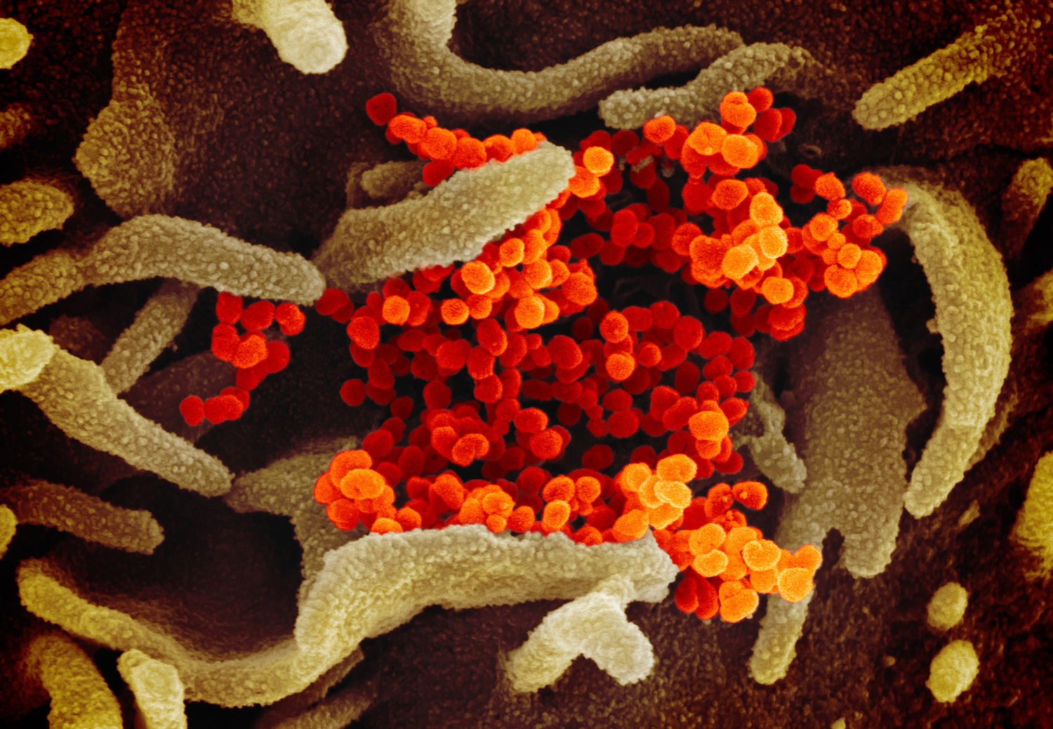
Investigational ChAdOx1 nCoV-19 vaccine protects monkeys against COVID-19  pneumonia | National Institutes of Health (NIH)
Investigational ChAdOx1 nCoV-19 vaccine protects monkeys against COVID-19 pneumonia | National Institutes of Health (NIH)
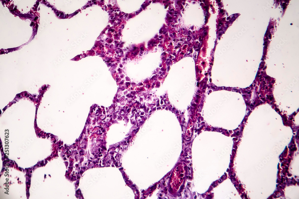
Nanoparticle Sensor Indicates Pneumonia Pathogen - BioTechniques

Histopathology of pneumonia, light micrograph, photo under microscope.  Cellulose aspiration pneumonia Stock Photo | Adobe Stock
Histopathology of pneumonia, light micrograph, photo under microscope. Cellulose aspiration pneumonia Stock Photo | Adobe Stock

Various bacteria cells in microscope. Streptococcus pneumonia,  pneumococcus, enterobacteriaceas, escherichia coli, salmonella, klebsiella  and others. Stock Illustration | Adobe Stock
Various bacteria cells in microscope. Streptococcus pneumonia, pneumococcus, enterobacteriaceas, escherichia coli, salmonella, klebsiella and others. Stock Illustration | Adobe Stock