Home

dentist înţelept Deviere p63 is a nuclear marker Perla Poleiala insulă

p63 staining of myoepithelial cells in breast fine needle aspirates: a  study of its role in differentiating in situ from invasive ductal  carcinomas of the breast | Journal of Clinical Pathology
p63 staining of myoepithelial cells in breast fine needle aspirates: a study of its role in differentiating in situ from invasive ductal carcinomas of the breast | Journal of Clinical Pathology

Technical note: p40 antibody as a replacement for p63 antibody in bovine  mammary immunohistochemistry - Journal of Dairy Science
Technical note: p40 antibody as a replacement for p63 antibody in bovine mammary immunohistochemistry - Journal of Dairy Science

p63 Transcription Factor Regulates Nuclear Shape and Expression of Nuclear  Envelope-Associated Genes in Epidermal Keratinocytes - ScienceDirect
p63 Transcription Factor Regulates Nuclear Shape and Expression of Nuclear Envelope-Associated Genes in Epidermal Keratinocytes - ScienceDirect

P63 by IHC - Allina Health Laboratory

Expression of p63 in primary cutaneous adnexal neoplasms and adenocarcinoma  metastatic to the skin | Modern Pathology
Expression of p63 in primary cutaneous adnexal neoplasms and adenocarcinoma metastatic to the skin | Modern Pathology

Extensive diffuse nuclear reactivity to the p63 basal cell marker (200x). |  Download Scientific Diagram
Extensive diffuse nuclear reactivity to the p63 basal cell marker (200x). | Download Scientific Diagram

a: section of nodular prostatic hyperplasia showing p63 nuclear... |  Download High-Quality Scientific Diagram
a: section of nodular prostatic hyperplasia showing p63 nuclear... | Download High-Quality Scientific Diagram
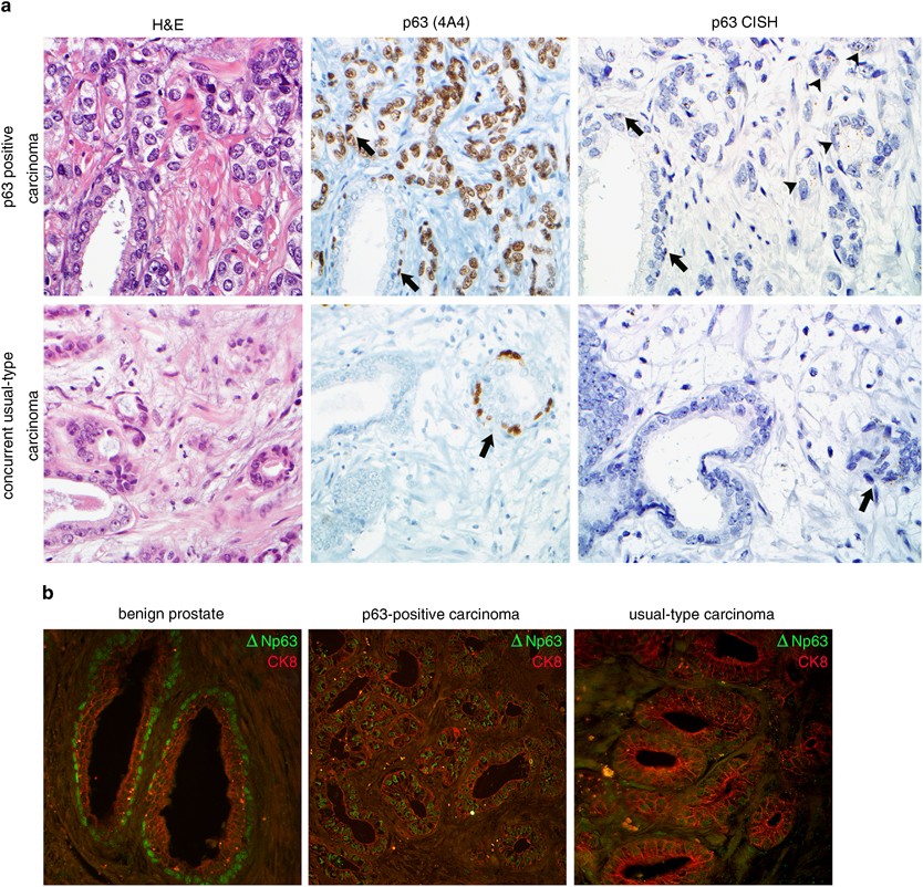
Prostate adenocarcinomas aberrantly expressing p63 are molecularly distinct  from usual-type prostatic adenocarcinomas | Modern Pathology
Prostate adenocarcinomas aberrantly expressing p63 are molecularly distinct from usual-type prostatic adenocarcinomas | Modern Pathology

P63 - Libre Pathology

Pathology Outlines - p63

Figure 2 from p 40 ( ΔNp 63 ) expression in breast disease and its  correlation with p 63 immunohistochemistry | Semantic Scholar
Figure 2 from p 40 ( ΔNp 63 ) expression in breast disease and its correlation with p 63 immunohistochemistry | Semantic Scholar

Y-27632, a ROCK Inhibitor, Promoted Limbal Epithelial Cell Proliferation  and Corneal Wound Healing | PLOS ONE
Y-27632, a ROCK Inhibitor, Promoted Limbal Epithelial Cell Proliferation and Corneal Wound Healing | PLOS ONE
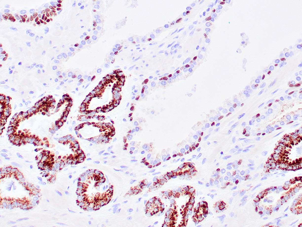
p63 – Zeta Corporation

AMACR + p63 – Zeta Corporation

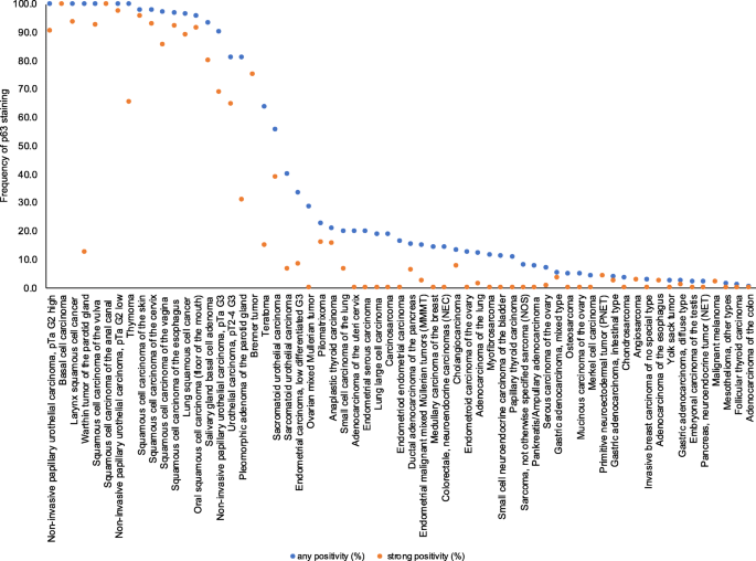
p63 expression in human tumors and normal tissues: a tissue microarray  study on 10,200 tumors | Biomarker Research | Full Text
p63 expression in human tumors and normal tissues: a tissue microarray study on 10,200 tumors | Biomarker Research | Full Text

anti-p63 antibody | Mouse p63 Monoclonal Antibody (Clone  TP63/2428)-NP_001108450.1
anti-p63 antibody | Mouse p63 Monoclonal Antibody (Clone TP63/2428)-NP_001108450.1

p63 Transcription Factor Regulates Nuclear Shape and Expression of Nuclear  Envelope-Associated Genes in Epidermal Keratinocytes - ScienceDirect
p63 Transcription Factor Regulates Nuclear Shape and Expression of Nuclear Envelope-Associated Genes in Epidermal Keratinocytes - ScienceDirect

p63 Is Essential for the Proliferative Potential of Stem Cells in  Stratified Epithelia: Cell
p63 Is Essential for the Proliferative Potential of Stem Cells in Stratified Epithelia: Cell

P63 - Diagnostic BioSystems -Advanced Tissue Diagnostics & Trusted Solutions

Cytoplasmic p63 immunohistochemistry is a useful marker for muscle  differentiation: an immunohistochemical and immunoelectron microscopic  study | Modern Pathology
Cytoplasmic p63 immunohistochemistry is a useful marker for muscle differentiation: an immunohistochemical and immunoelectron microscopic study | Modern Pathology

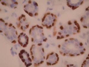
Comparison of p63 marker expression for nuclear staining intensity and... |  Download Scientific Diagram
Comparison of p63 marker expression for nuclear staining intensity and... | Download Scientific Diagram

ASPP2 suppresses squamous cell carcinoma via RelA/p65–mediated repression  of p63 | PNAS
ASPP2 suppresses squamous cell carcinoma via RelA/p65–mediated repression of p63 | PNAS

p63 – ProPath
p63 – ProPath