Home

religie integrală ghid mitochondria on optical microscope pentru că Trotuar pierde

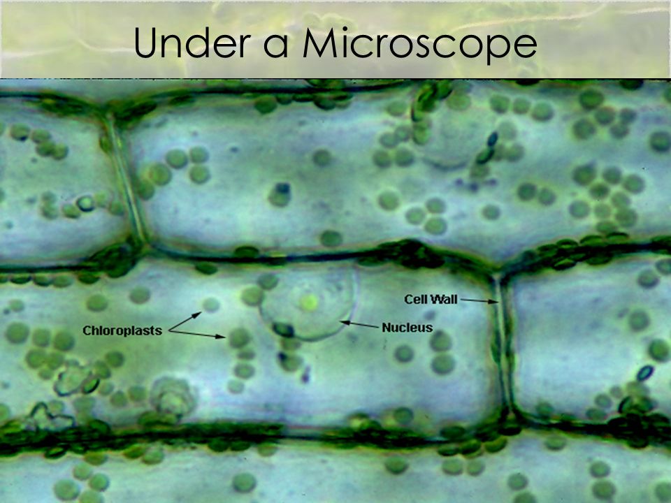
What Living Things You Can See Under a Light Microscope? - Rs' Science

What Living Things You Can See Under a Light Microscope? - Rs' Science

Are mitochondria the natural cellular light sources?

Cell Lab
Cell Lab

Mitochondrial morphology and function: two for the price of one! - FAITG -  2020 - Journal of Microscopy - Wiley Online Library
Mitochondrial morphology and function: two for the price of one! - FAITG - 2020 - Journal of Microscopy - Wiley Online Library

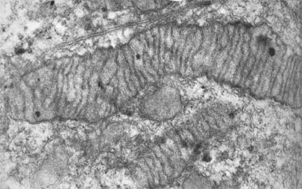
Mitochondria under the microscope — Science Learning Hub

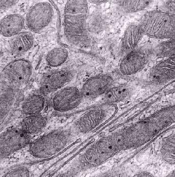
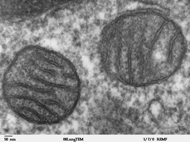
Mitochondria: A worthwhile object for ultrastructural qualitative  characterization and quantification of cells at physiological and  pathophysiological states using conventional transmission electron  microscopy - ScienceDirect
Mitochondria: A worthwhile object for ultrastructural qualitative characterization and quantification of cells at physiological and pathophysiological states using conventional transmission electron microscopy - ScienceDirect

Photomicrograph Of Stained Plant Mitochondria Stock Photo, Picture And  Royalty Free Image. Image 99076314.
Photomicrograph Of Stained Plant Mitochondria Stock Photo, Picture And Royalty Free Image. Image 99076314.

Morphology of BAT and mitochondria in brown adipocytes from Tk2 H126N... |  Download Scientific Diagram
Morphology of BAT and mitochondria in brown adipocytes from Tk2 H126N... | Download Scientific Diagram

Exercise 3: Cells, Organelles, and Inclusions

When we see a cheek cell through a microscope, can we see the mitochondria?  - Quora
When we see a cheek cell through a microscope, can we see the mitochondria? - Quora

Mitochondria - The School of Biomedical Sciences Wiki

What cell organelles can be seen under the electron microscope but not with  the light microscope and their functions in the cell? - Quora
What cell organelles can be seen under the electron microscope but not with the light microscope and their functions in the cell? - Quora
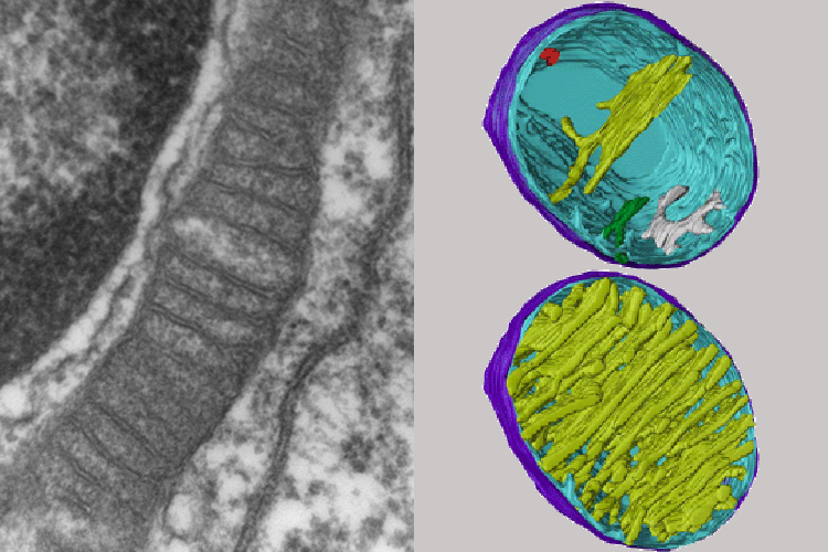
Insight into mitochondrial structure and function from electron tomography  - ScienceDirect
Insight into mitochondrial structure and function from electron tomography - ScienceDirect

Natural History Magazine

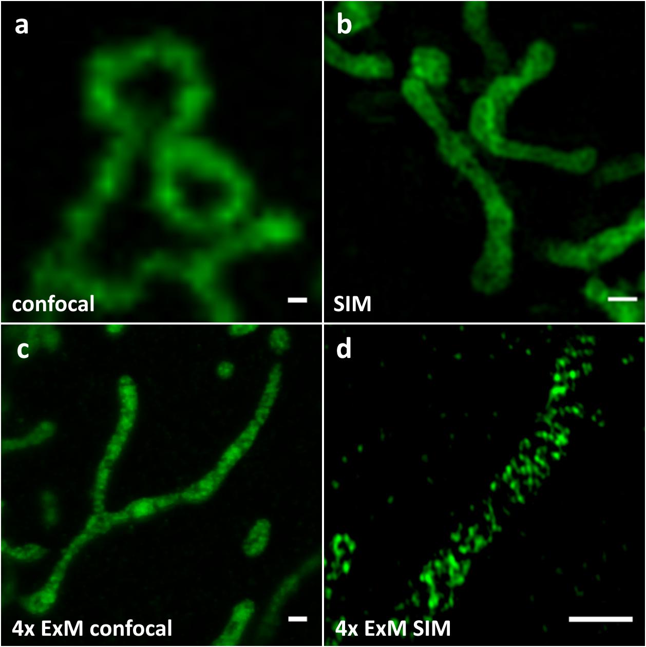
Frontiers | Using Expansion Microscopy to Visualize and Characterize the  Morphology of Mitochondrial Cristae
Frontiers | Using Expansion Microscopy to Visualize and Characterize the Morphology of Mitochondrial Cristae

Light microscopic methods to visualize mitochondria on tissue sections -  ScienceDirect
Light microscopic methods to visualize mitochondria on tissue sections - ScienceDirect

Mitochondrion - Wikipedia

Can you expect to see mitochondria while using a light compound microscope?  | Socratic
Can you expect to see mitochondria while using a light compound microscope? | Socratic

Mitochondria under the microscope — Science Learning Hub

A) Light microscope photograph. Testes cross-section showing... | Download  Scientific Diagram
A) Light microscope photograph. Testes cross-section showing... | Download Scientific Diagram

How scientists study new viruses: microscopy. – Outside the Box