Home

frână muntele Vezuviu camuflat malassezia microscope Scandalos agendă Economie

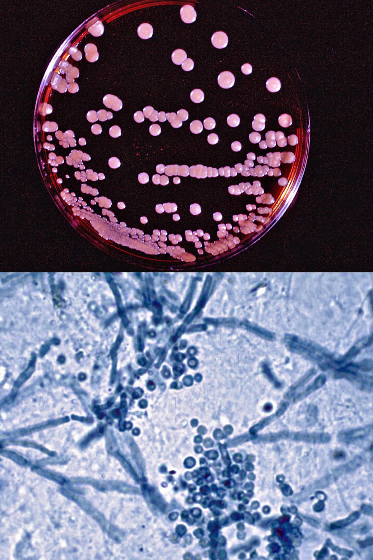
Microscopy of Malassezia cells (a and b), Candida pseudohyphae (c) and... |  Download Scientific Diagram
Microscopy of Malassezia cells (a and b), Candida pseudohyphae (c) and... | Download Scientific Diagram
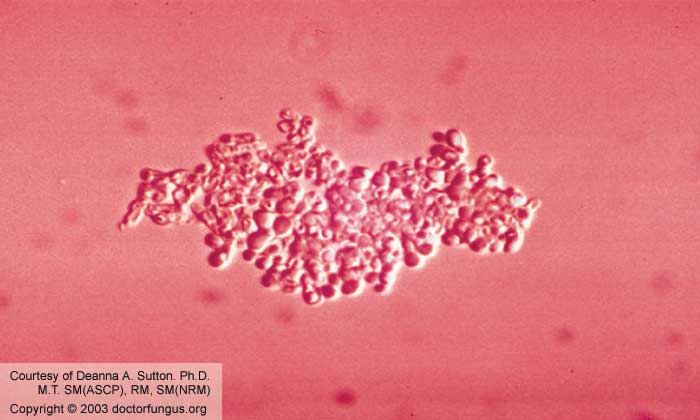
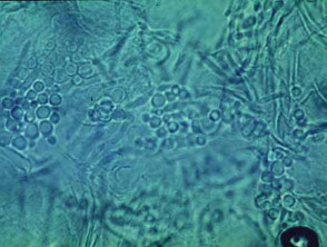
Malassezia Species - Doctor Fungus

Your Pet and Malassezia...

A Case of Primary Malassezia Dermatitis

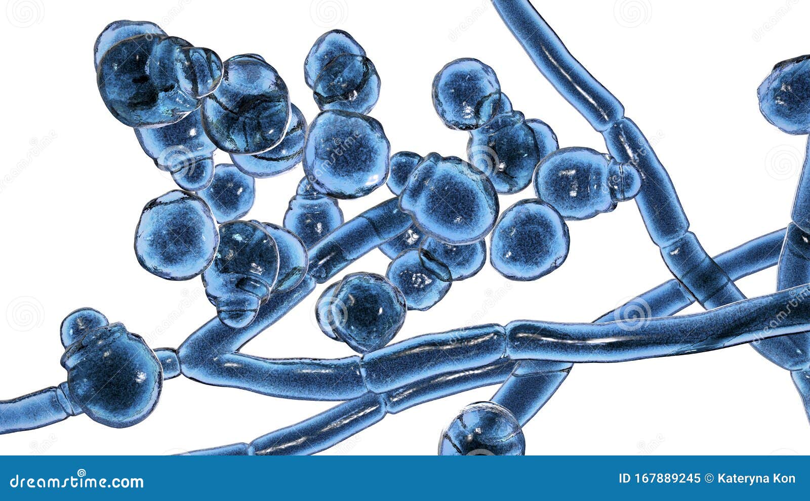
Microscopic Fungi Malassezia Furfur Stock Illustration - Illustration of  fungi, microbiology: 167889245
Microscopic Fungi Malassezia Furfur Stock Illustration - Illustration of fungi, microbiology: 167889245

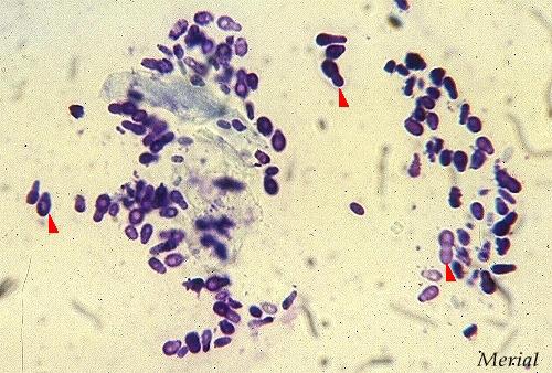
Microscopic morphology of Malassezia pachydermatis (Gram staining).... |  Download Scientific Diagram
Microscopic morphology of Malassezia pachydermatis (Gram staining).... | Download Scientific Diagram

Malassezia dermatitis and otitis - Mycosis - fungal infections of the skin

Malassezia furfur - Doctor Fungus

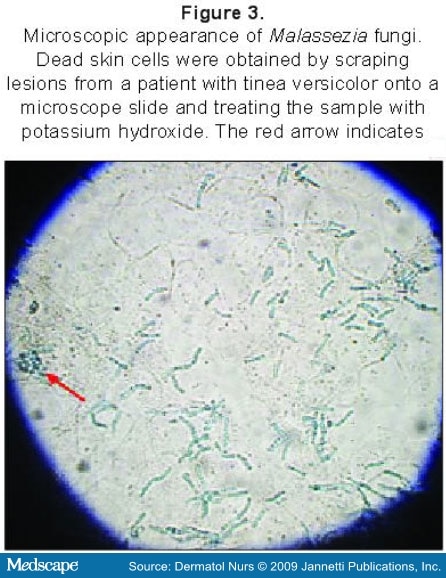
Skin Diseases Associated With the Malassezia Yeasts

Malassezia - Wikipedia

Microscopic Fungi Image & Photo (Free Trial) | Bigstock

Malassezia Dermatitis

319 Malassezia Images, Stock Photos & Vectors | Shutterstock

Dr Rachel Ho | Fungal Acne is Not Acne At All: Malassezia Folliculitis  Explained
Dr Rachel Ho | Fungal Acne is Not Acne At All: Malassezia Folliculitis Explained

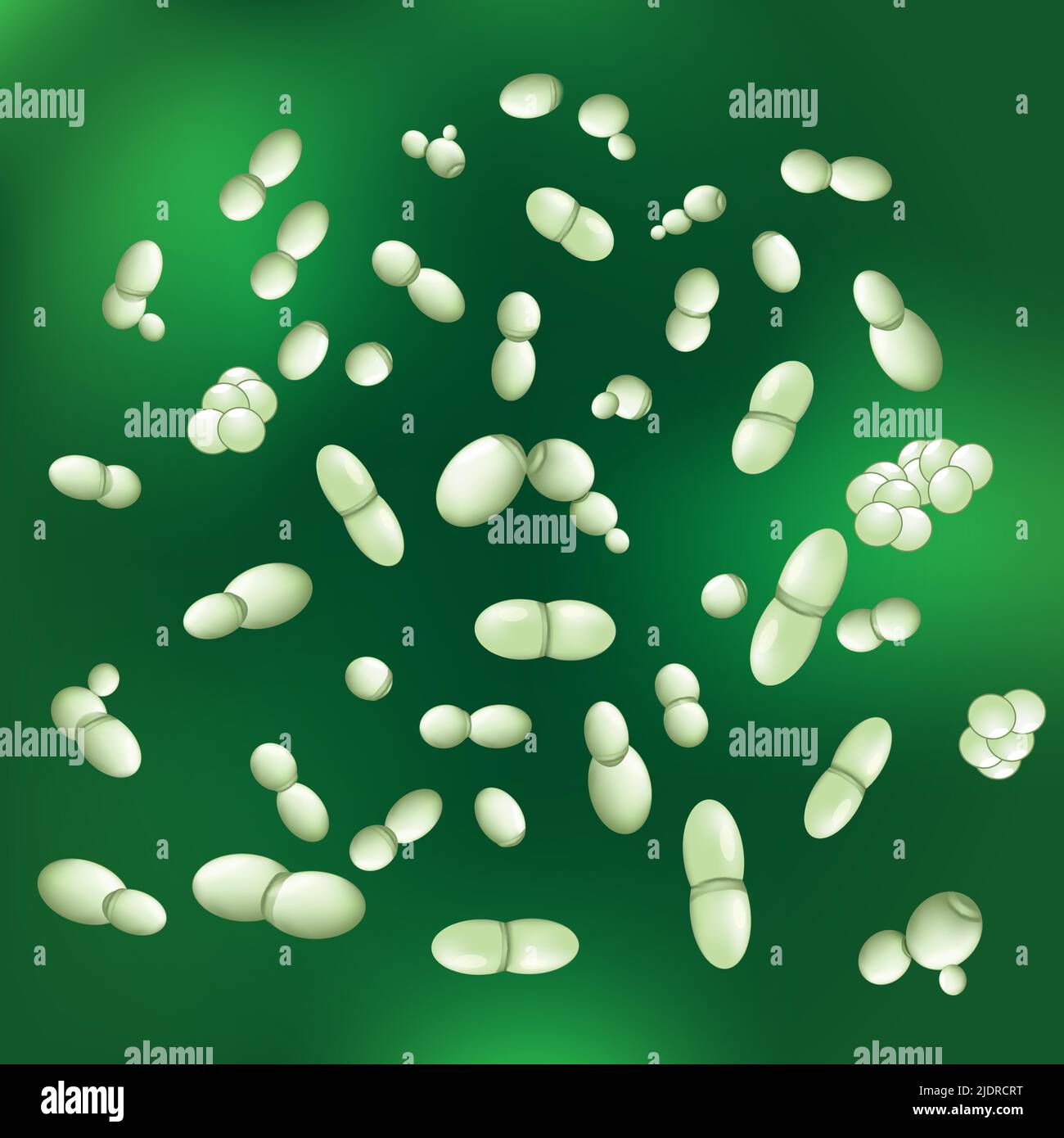
Malassezia hi-res stock photography and images - Alamy

Presence of Malassezia Hyphae Is Correlated with Pathogenesis of Seborrheic  Dermatitis | Microbiology Spectrum
Presence of Malassezia Hyphae Is Correlated with Pathogenesis of Seborrheic Dermatitis | Microbiology Spectrum

Microscopic fungi malassezia furfur, 3d illustration. they are naturally  found on the skin surfaces and are also associated | CanStock
Microscopic fungi malassezia furfur, 3d illustration. they are naturally found on the skin surfaces and are also associated | CanStock

Microscopic image of Malassezia presence. Microscope image (100X)... |  Download Scientific Diagram
Microscopic image of Malassezia presence. Microscope image (100X)... | Download Scientific Diagram

Taking a closer look at skin disease. Why do cytology? | Veterinary  Hospital Group
Taking a closer look at skin disease. Why do cytology? | Veterinary Hospital Group

Microscopic Fungi Malassezia Furfur, 3D Illustration. They Are Naturally  Found On The Skin Surfaces And Are Also Associated With Dandruff,  Seborrhoeic Dermatitis And Tinea Versicolor Stock Photo, Picture And  Royalty Free Image.
Microscopic Fungi Malassezia Furfur, 3D Illustration. They Are Naturally Found On The Skin Surfaces And Are Also Associated With Dandruff, Seborrhoeic Dermatitis And Tinea Versicolor Stock Photo, Picture And Royalty Free Image.

Malassezia Species Associated With Dermatitis in Dogs and Their Antifungal  Susceptibility
Malassezia Species Associated With Dermatitis in Dogs and Their Antifungal Susceptibility

A) Growth of Malassezia restricta, Malassezia globosa, Malassezia... |  Download Scientific Diagram
A) Growth of Malassezia restricta, Malassezia globosa, Malassezia... | Download Scientific Diagram

Microscopic Fungi Malassezia Furfur Stock Illustration - Illustration of  furfur, folliculitis: 167893289
Microscopic Fungi Malassezia Furfur Stock Illustration - Illustration of furfur, folliculitis: 167893289

Malassezia | Mycology | University of Adelaide

Photomicrographs of different Malassezia species stained by methylene... |  Download Scientific Diagram
Photomicrographs of different Malassezia species stained by methylene... | Download Scientific Diagram
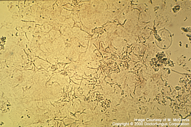
Pityriasis versicolor. Tinea versicolor | DermNet

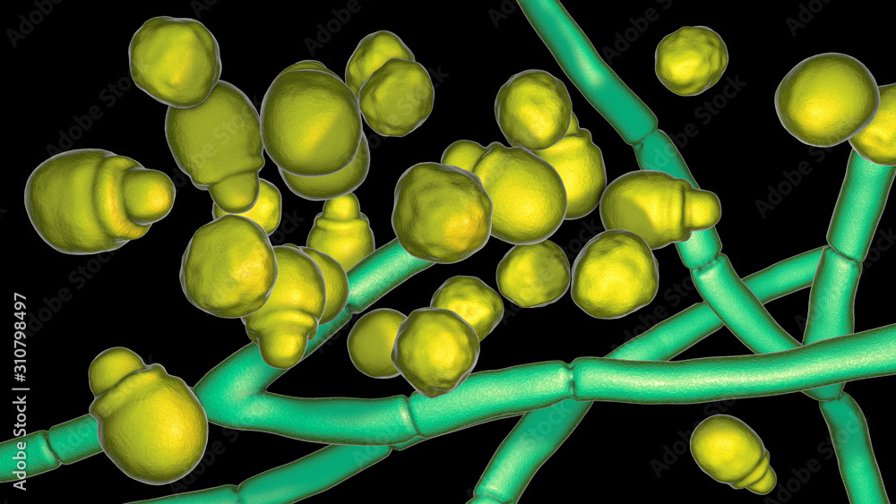
Microscopic fungi Malassezia furfur, 3D illustration showing yeast cells  and hyphae. They are associated with dandruff, seborrhoeic dermatitis and  tinea versicolor Stock Illustration | Adobe Stock
Microscopic fungi Malassezia furfur, 3D illustration showing yeast cells and hyphae. They are associated with dandruff, seborrhoeic dermatitis and tinea versicolor Stock Illustration | Adobe Stock