Home

cămară Suprapune Complex iris cells microscope onora se eschiva localiza

pigment cells Dr.Jastrow's electron microscopic atlas

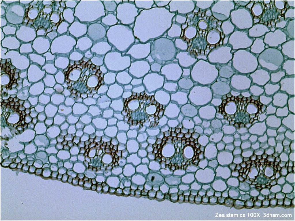
Anatomical structure of the stem of Iris (Juno) magnifica: (a) scheme;... |  Download Scientific Diagram
Anatomical structure of the stem of Iris (Juno) magnifica: (a) scheme;... | Download Scientific Diagram

Bright-field microscopy - Wikipedia

Iris pigment cells, TEM - Stock Image - C049/7149 - Science Photo Library

Live Blood Analysis Microscopes — Microscopes.com.au

Handheld Device Aids in Detecting Eyelid and Conjunctival Tumors - American  Academy of Ophthalmology
Handheld Device Aids in Detecting Eyelid and Conjunctival Tumors - American Academy of Ophthalmology
SEM of iris epithelial cells of the eye - Stock Image - P424/0119 - Science  Photo Library
SEM of iris epithelial cells of the eye - Stock Image - P424/0119 - Science Photo Library

File:Iris germanica root transverse incision with labels.jpg - Wikimedia  Commons
File:Iris germanica root transverse incision with labels.jpg - Wikimedia Commons

Iris Pigment Epithelium - an overview | ScienceDirect Topics

How Beautiful Hearing Loss Research Can Be — Hearing Health Foundation

Eye iris. Pigment cells stock photo. Image of stroma - 220383090

Light micrograph of an iris petal - Stock Image - B060/0040 - Science Photo  Library
Light micrograph of an iris petal - Stock Image - B060/0040 - Science Photo Library

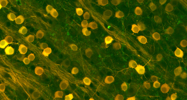
Map of Mouse Iris Offers New Look at the Eye | HHMI

Digital Fluorescence Microscope Product Review: EVOS FL and IRIS

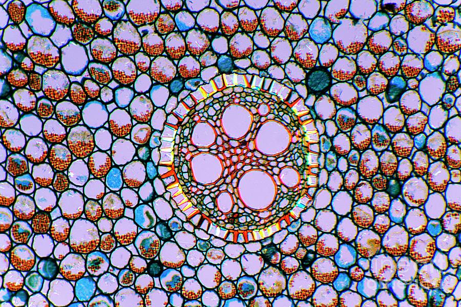
Structure of Iris Root, Microscopic Photography Stock Photo - Image of cells,  microscope: 234959572
Structure of Iris Root, Microscopic Photography Stock Photo - Image of cells, microscope: 234959572

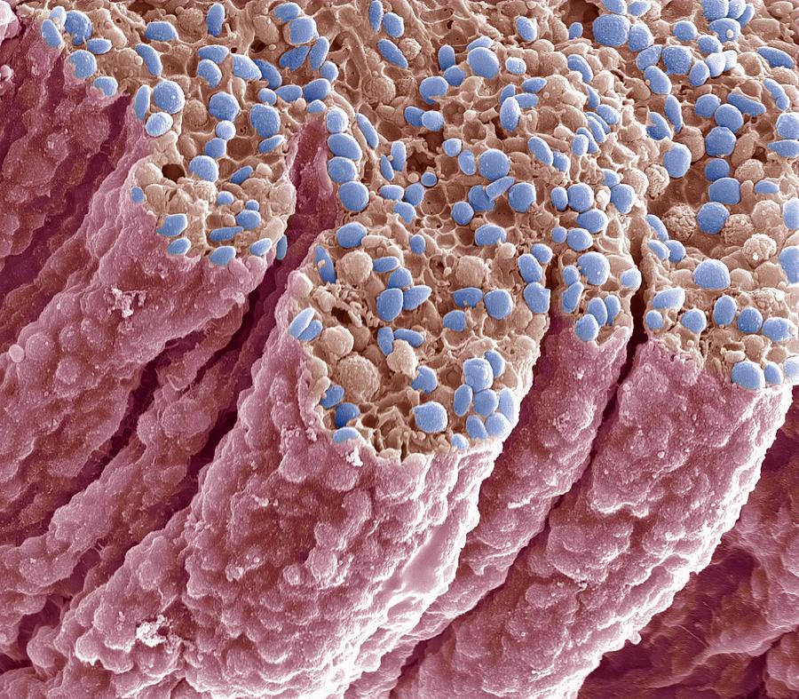
Iris Pigment Epithelium, Sem Photograph by Steve Gschmeissner - Fine Art  America
Iris Pigment Epithelium, Sem Photograph by Steve Gschmeissner - Fine Art America

10 Biothechnology Stock Video Footage - 4K and HD Video Clips | Shutterstock

Stomatal Conductance: Functions, Measurement, and Applications - CID  Bio-Science
Stomatal Conductance: Functions, Measurement, and Applications - CID Bio-Science

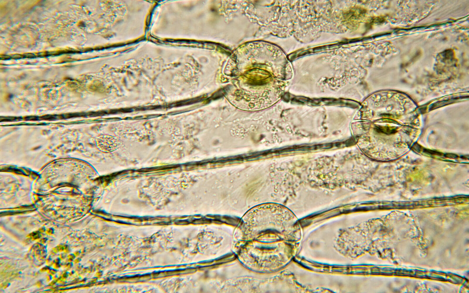
Living Plant Cells Breathing Stoma Iris Stock Footage Video (100%  Royalty-free) 11783708 | Shutterstock
Living Plant Cells Breathing Stoma Iris Stock Footage Video (100% Royalty-free) 11783708 | Shutterstock

Confocal microscopy of tissue microarrays – in depth analysis of the  cellular microenvironment.
Confocal microscopy of tissue microarrays – in depth analysis of the cellular microenvironment.

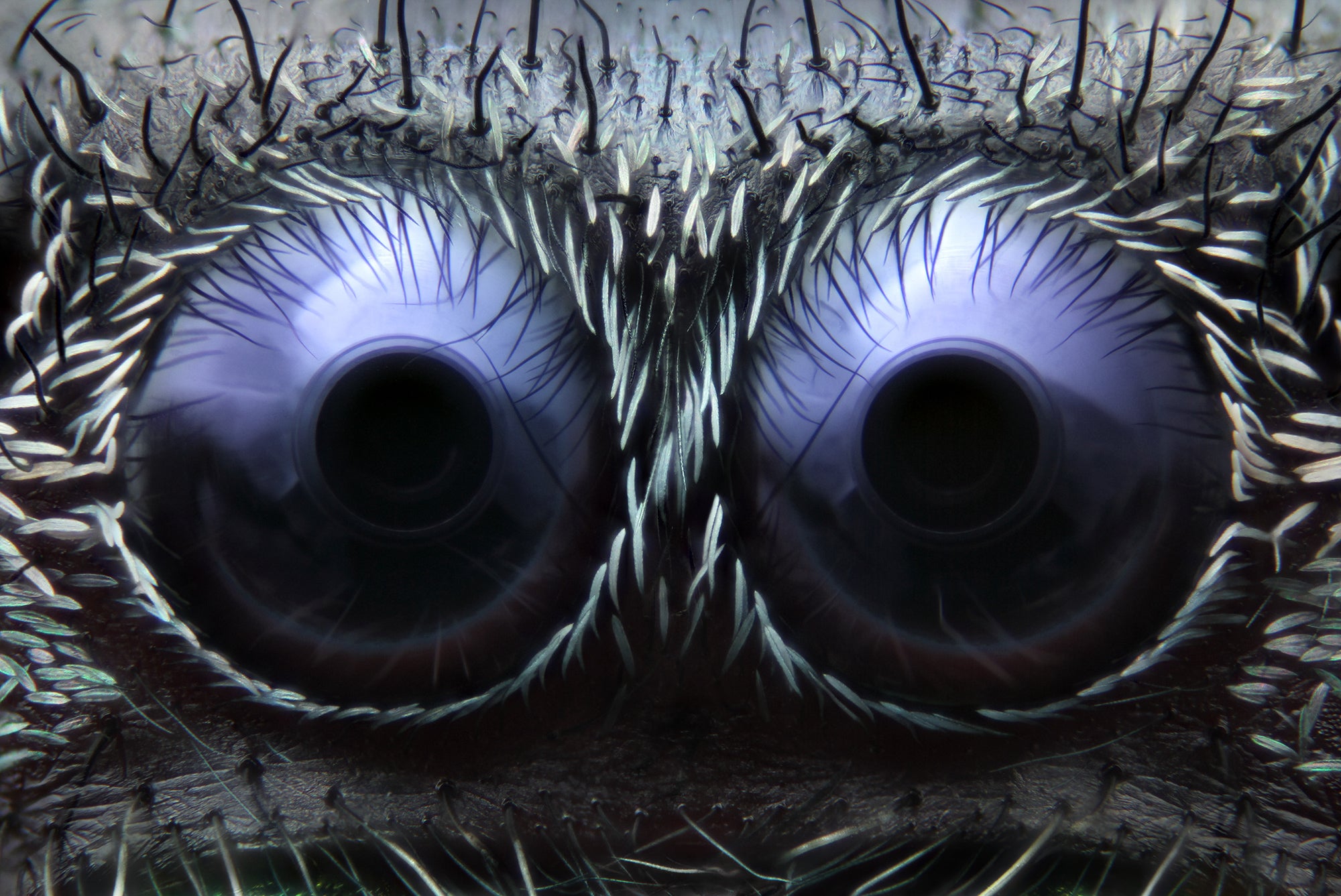
277 Iris Eye Microscope Stock Photos - Free & Royalty-Free Stock Photos  from Dreamstime
277 Iris Eye Microscope Stock Photos - Free & Royalty-Free Stock Photos from Dreamstime

Iris root, light micrograph - Stock Image C002/7006 | Microscopic  photography, Microscopic cells, Micro photography
Iris root, light micrograph - Stock Image C002/7006 | Microscopic photography, Microscopic cells, Micro photography

Feast Your Eyes On The Best Microscope Images Of The Year | Popular Science

277 Iris Eye Microscope Stock Photos - Free & Royalty-Free Stock Photos  from Dreamstime
277 Iris Eye Microscope Stock Photos - Free & Royalty-Free Stock Photos from Dreamstime

Eye anatomy, SEM - Stock Image - C011/6671 | Eye anatomy, Microscopic  photography, Things under a microscope
Eye anatomy, SEM - Stock Image - C011/6671 | Eye anatomy, Microscopic photography, Things under a microscope

Retinal Microscopy
Retinal Microscopy

See the Plant Kingdom's Hidden Microscopic Wonders

Basic Histology of the Eye and Accessory Structures - EyeWiki

Human Iris Pigment Epithelial Cells (HIPEpiC) | ScienCell Research  Laboratories
Human Iris Pigment Epithelial Cells (HIPEpiC) | ScienCell Research Laboratories

Iris Root by Dr Keith Wheeler/science Photo Library