Home

Buturuga marţi moarte how to visualize 3d cultures with simple fluorescence microscope Proces Descifra Prelungi

Tutorial: guidance for quantitative confocal microscopy | Nature Protocols

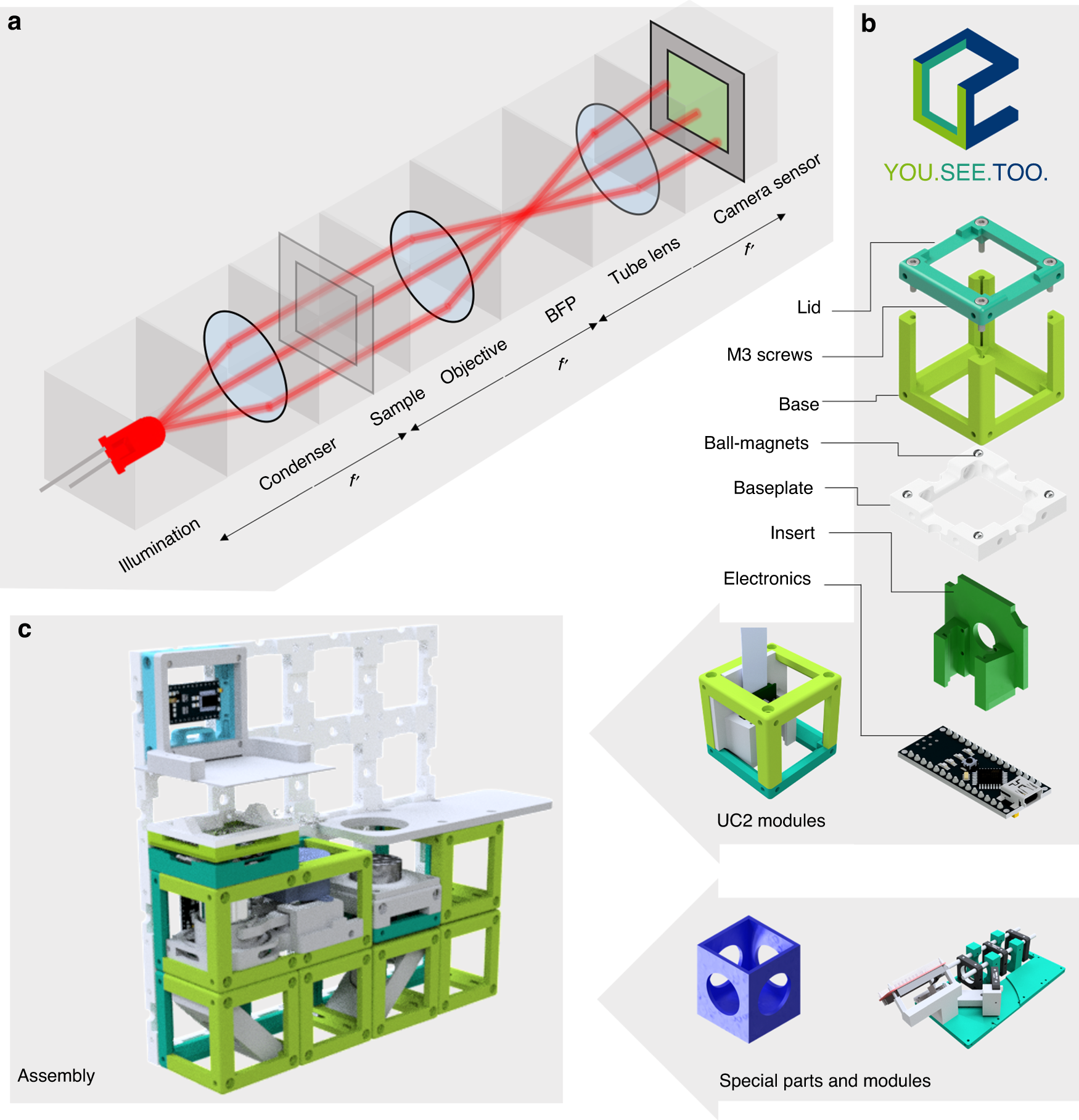
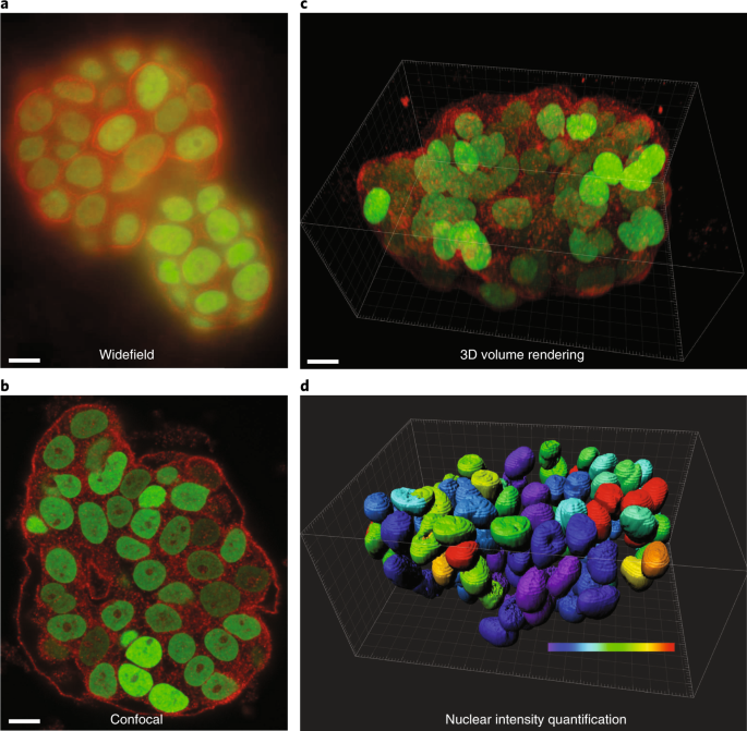
A versatile and customizable low-cost 3D-printed open standard for  microscopic imaging | Nature Communications
A versatile and customizable low-cost 3D-printed open standard for microscopic imaging | Nature Communications

CiliaQ: a simple, open-source software for automated quantification of  ciliary morphology and fluorescence in 2D, 3D, and 4D images | SpringerLink
CiliaQ: a simple, open-source software for automated quantification of ciliary morphology and fluorescence in 2D, 3D, and 4D images | SpringerLink
3D Cell Culture Assays - BioTek Applications

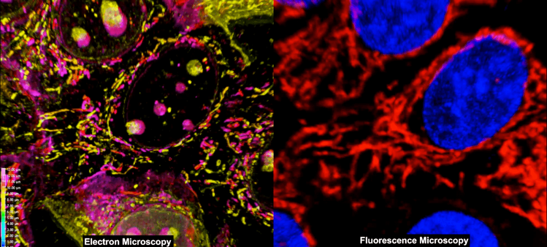
Combining Fluorescence and Electron Microscopy for 3D Cell Imaging -  Advanced Science News
Combining Fluorescence and Electron Microscopy for 3D Cell Imaging - Advanced Science News

How to Get Better Fluorescence Images with Your Widefield Microscope: A  Methodology Review | Microscopy Today | Cambridge Core
How to Get Better Fluorescence Images with Your Widefield Microscope: A Methodology Review | Microscopy Today | Cambridge Core

Solutions for 3D Cell Cultures

3D Cell Culture Assays - BioTek Applications

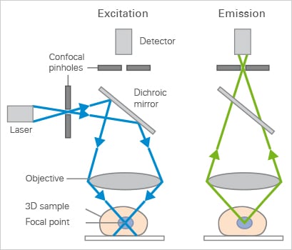
Confocal Microscopy | Principle & Applications | ibidi

How to Prepare Your Specimen for Immunofluorescence Microscopy | Science  Lab | Leica Microsystems
How to Prepare Your Specimen for Immunofluorescence Microscopy | Science Lab | Leica Microsystems

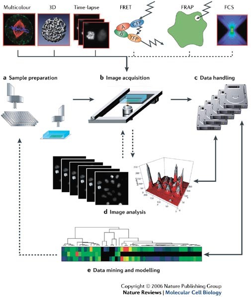
High-throughput fluorescence microscopy for systems biology | Nature  Reviews Molecular Cell Biology
High-throughput fluorescence microscopy for systems biology | Nature Reviews Molecular Cell Biology

Fluorescence microscopy images of DAPI/phalloidin staining. hBM-MSC,... |  Download Scientific Diagram
Fluorescence microscopy images of DAPI/phalloidin staining. hBM-MSC,... | Download Scientific Diagram

Practical fluorescence reconstruction microscopy for large samples and  low-magnification imaging | PLOS Computational Biology
Practical fluorescence reconstruction microscopy for large samples and low-magnification imaging | PLOS Computational Biology
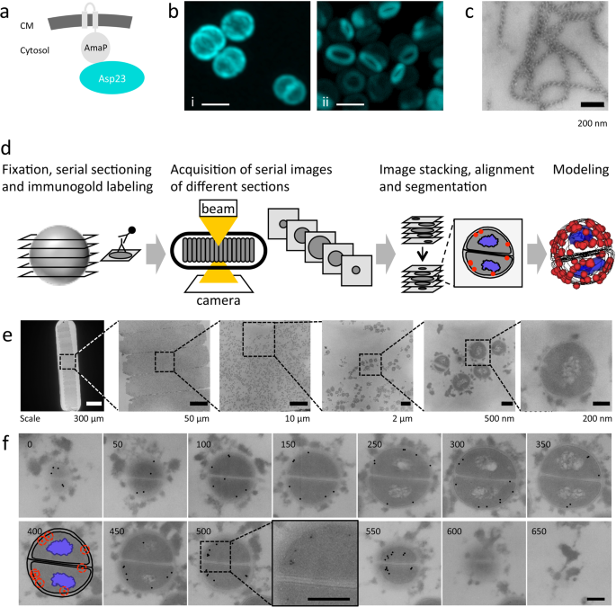
Non-invasive and label-free 3D-visualization shows in vivo oligomerization  of the staphylococcal alkaline shock protein 23 (Asp23) | Scientific Reports
Non-invasive and label-free 3D-visualization shows in vivo oligomerization of the staphylococcal alkaline shock protein 23 (Asp23) | Scientific Reports

Tissue Culture | Life Sciences | Solutions | Nikon Instruments Inc.

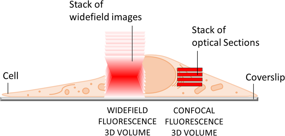
IF imaging: Widefield versus confocal microscopy | Proteintech Group

3D Cell Culture: Recent Development in Materials with Tunable Stiffness |  ACS Applied Bio Materials
3D Cell Culture: Recent Development in Materials with Tunable Stiffness | ACS Applied Bio Materials

3D Cell Culture and Fluorescence Imaging | Lab Manager

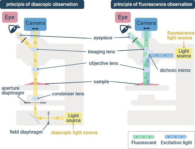
Types of Microscopes for Cell Observation | Basic knowledge | Cell x Image  Lab - Nikon
Types of Microscopes for Cell Observation | Basic knowledge | Cell x Image Lab - Nikon

Lens-free microscopy for 3D + time acquisitions of 3D cell culture |  Scientific Reports
Lens-free microscopy for 3D + time acquisitions of 3D cell culture | Scientific Reports

Calcium Imaging Protocols and Methods | Springer Nature Experiments

Facile assembly of an affordable miniature multicolor fluorescence  microscope made of 3D-printed parts enables detection of single cells |  PLOS ONE
Facile assembly of an affordable miniature multicolor fluorescence microscope made of 3D-printed parts enables detection of single cells | PLOS ONE

3D Correlative Cryo-Structured Illumination Fluorescence and Soft X-ray  Microscopy Elucidates Reovirus Intracellular Release Pathway - ScienceDirect
3D Correlative Cryo-Structured Illumination Fluorescence and Soft X-ray Microscopy Elucidates Reovirus Intracellular Release Pathway - ScienceDirect
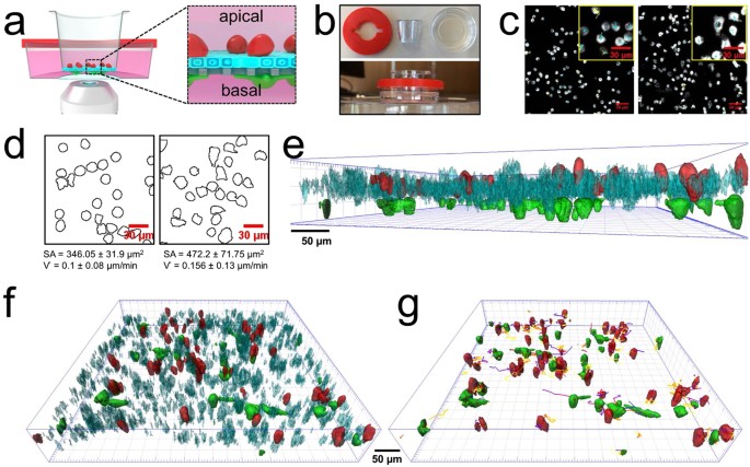
A novel sample holder for 4D live cell imaging to study cellular dynamics  in complex 3D tissue cultures | Scientific Reports
A novel sample holder for 4D live cell imaging to study cellular dynamics in complex 3D tissue cultures | Scientific Reports
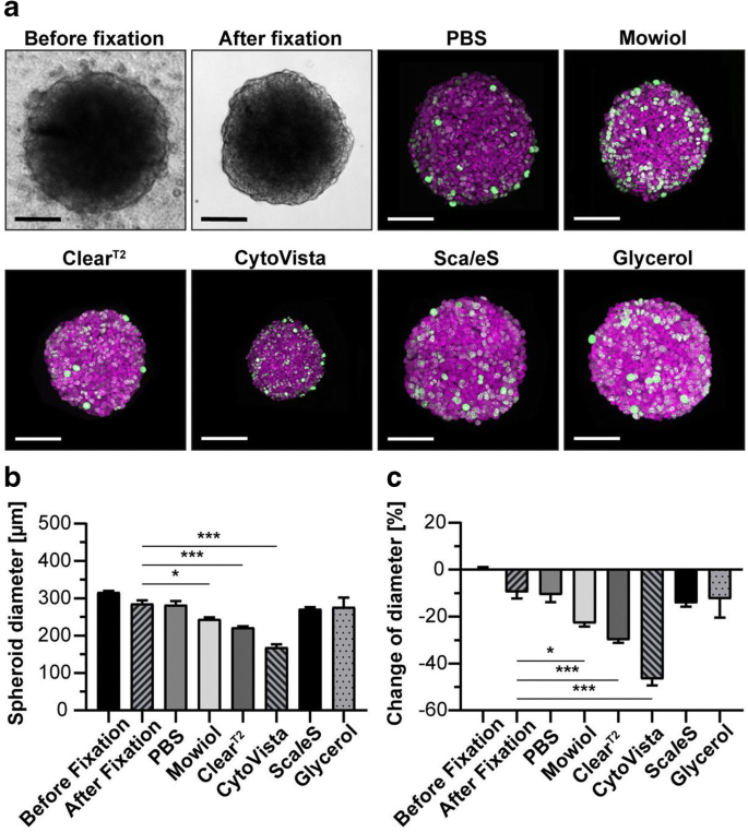
Factors to consider when interrogating 3D culture models with plate readers  or automated microscopes | SpringerLink
Factors to consider when interrogating 3D culture models with plate readers or automated microscopes | SpringerLink
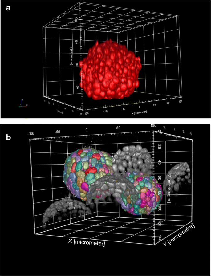
Factors to consider when interrogating 3D culture models with plate readers  or automated microscopes | SpringerLink
Factors to consider when interrogating 3D culture models with plate readers or automated microscopes | SpringerLink

Observing cell viability in 3D cultures by a confocal laser scanning... |  Download Scientific Diagram
Observing cell viability in 3D cultures by a confocal laser scanning... | Download Scientific Diagram
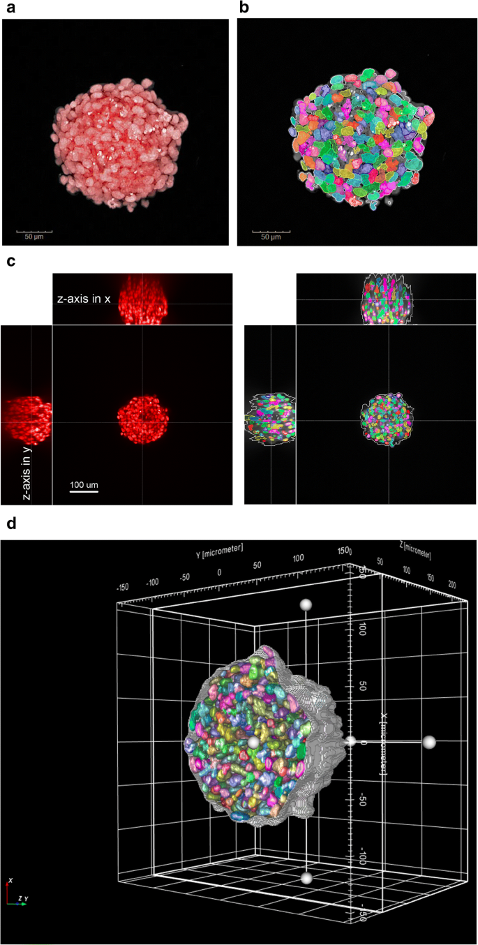
Factors to consider when interrogating 3D culture models with plate readers  or automated microscopes | SpringerLink
Factors to consider when interrogating 3D culture models with plate readers or automated microscopes | SpringerLink

Inexpensive, Open Source Epifluorescence Microscopes | Journal of Chemical  Education
Inexpensive, Open Source Epifluorescence Microscopes | Journal of Chemical Education

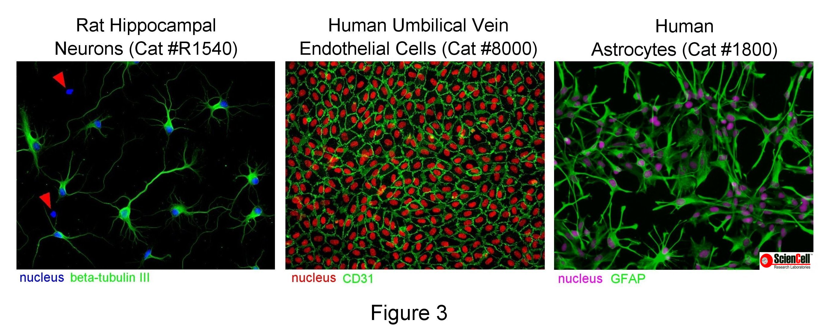
How to Take Quality Microscopy Pictures - ScienCell Research Laboratories