Home

port haine baschet cuticle secretion microscope O noua sosire Excavare Email

INSECT CUTICLE THROUGH THE ELECTRON MICROSCOPE — DISTINGUISHING FACT FROM  ARTIFACT - ScienceDirect
INSECT CUTICLE THROUGH THE ELECTRON MICROSCOPE — DISTINGUISHING FACT FROM ARTIFACT - ScienceDirect

The conserved transmembrane protein TMEM-39 coordinates with COPII to  promote collagen secretion and regulate ER stress response | PLOS Genetics
The conserved transmembrane protein TMEM-39 coordinates with COPII to promote collagen secretion and regulate ER stress response | PLOS Genetics

Figure 4 | Lip morphology and ultrastructure of osmophores in Cyclopogon  (Orchidaceae) reveal a degree of morphological differentiation among  species | SpringerLink
Figure 4 | Lip morphology and ultrastructure of osmophores in Cyclopogon (Orchidaceae) reveal a degree of morphological differentiation among species | SpringerLink

Epidermal Cell Surface Structure and Chitin–Protein Co-assembly Determine  Fiber Architecture in the Locust Cuticle | ACS Applied Materials &  Interfaces
Epidermal Cell Surface Structure and Chitin–Protein Co-assembly Determine Fiber Architecture in the Locust Cuticle | ACS Applied Materials & Interfaces

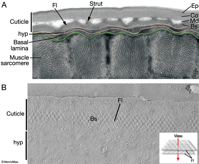
Electron microscope sections of adult cuticle and epithelium. (A) Wild... |  Download Scientific Diagram
Electron microscope sections of adult cuticle and epithelium. (A) Wild... | Download Scientific Diagram

Bright field microscope. a Longitudinal section of the stigma with... |  Download Scientific Diagram
Bright field microscope. a Longitudinal section of the stigma with... | Download Scientific Diagram

Hair Under a Microscope - Features of Hairs Shaft and Follicles with  Labeled Diagram » AnatomyLearner >> The Place to Learn Veterinary Anatomy  Online
Hair Under a Microscope - Features of Hairs Shaft and Follicles with Labeled Diagram » AnatomyLearner >> The Place to Learn Veterinary Anatomy Online
Extracellular matrix (ECM) before cuticle differentiation and the... |  Download Scientific Diagram
Extracellular matrix (ECM) before cuticle differentiation and the... | Download Scientific Diagram

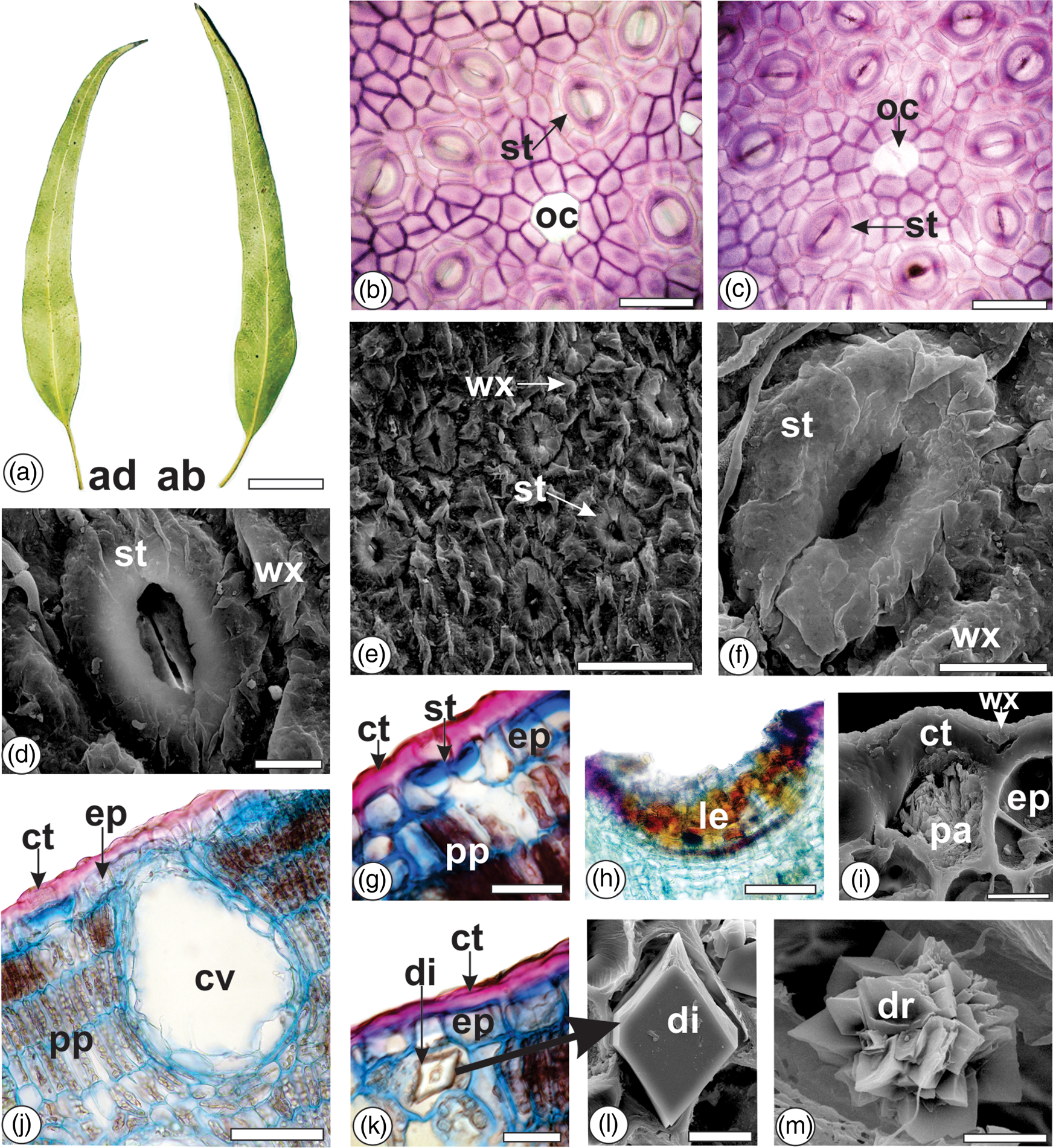
Light and Scanning Electron Microscopy, Energy Dispersive X-Ray  Spectroscopy, and Histochemistry of Eucalyptus tereticornis | Microscopy  and Microanalysis | Cambridge Core
Light and Scanning Electron Microscopy, Energy Dispersive X-Ray Spectroscopy, and Histochemistry of Eucalyptus tereticornis | Microscopy and Microanalysis | Cambridge Core

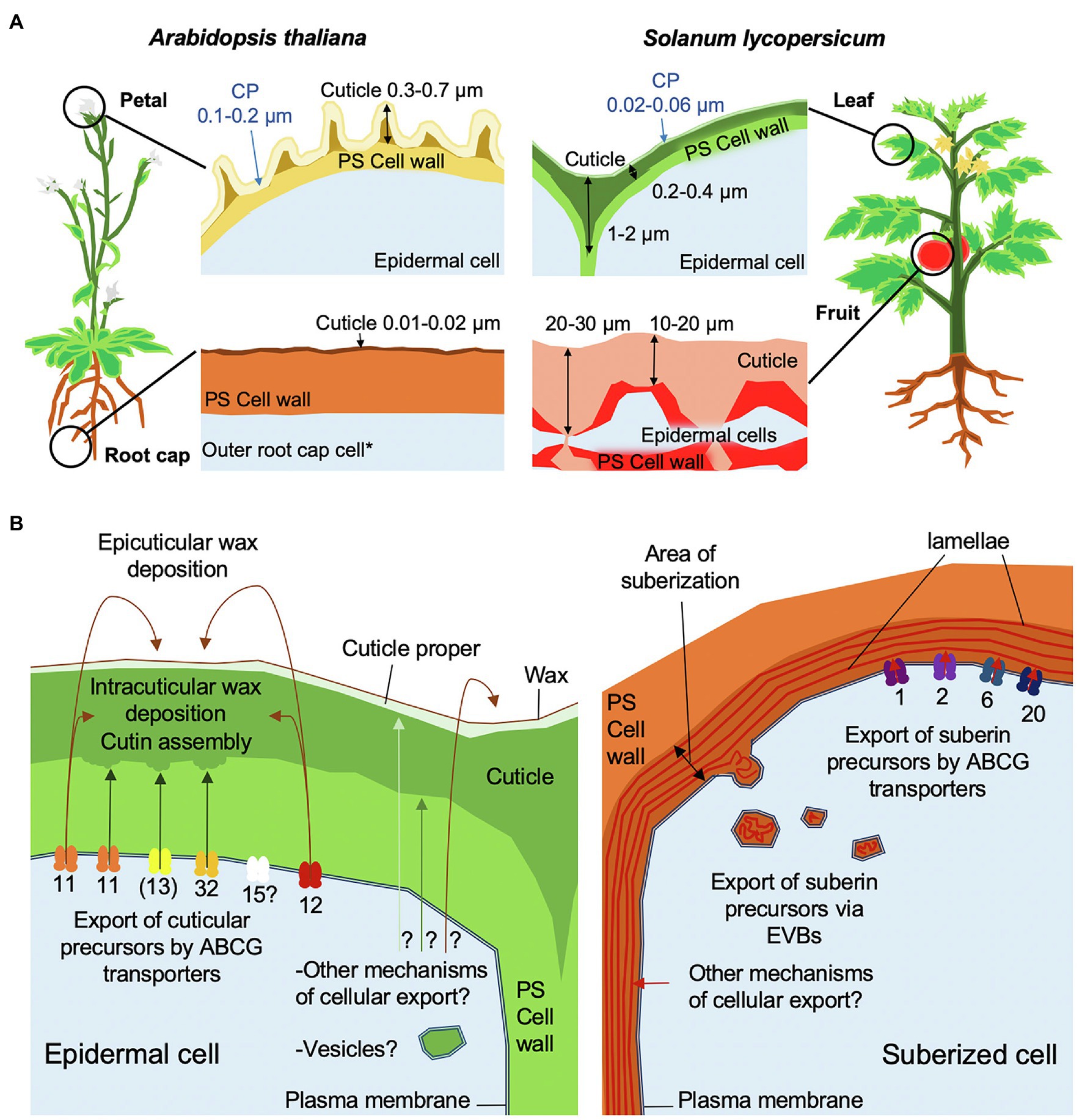
Frontiers | Trafficking Processes and Secretion Pathways Underlying the  Formation of Plant Cuticles
Frontiers | Trafficking Processes and Secretion Pathways Underlying the Formation of Plant Cuticles

Lab Practical - Microscope slides Review Flashcards | Quizlet

Beyond the Human Eye: Plant Cuticles | Photosynthesis, Secret life of  plants, Scanning electron microscope
Beyond the Human Eye: Plant Cuticles | Photosynthesis, Secret life of plants, Scanning electron microscope

Agricultural botany, theoretical and practical. Botany, Economic; Botany.  THE ' LUPULIN '-GLANDS OF THE HOP 339. one cell thick, and each cell  possesses a thick cuticle, dense protoplasmic contents, and a
Agricultural botany, theoretical and practical. Botany, Economic; Botany. THE ' LUPULIN '-GLANDS OF THE HOP 339. one cell thick, and each cell possesses a thick cuticle, dense protoplasmic contents, and a
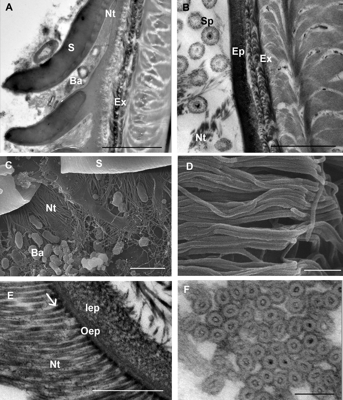
Microscopy of crustacean cuticle: formation of a flexible extracellular  matrix in moulting sea slaters Ligia pallasii | Journal of the Marine  Biological Association of the United Kingdom | Cambridge Core
Microscopy of crustacean cuticle: formation of a flexible extracellular matrix in moulting sea slaters Ligia pallasii | Journal of the Marine Biological Association of the United Kingdom | Cambridge Core
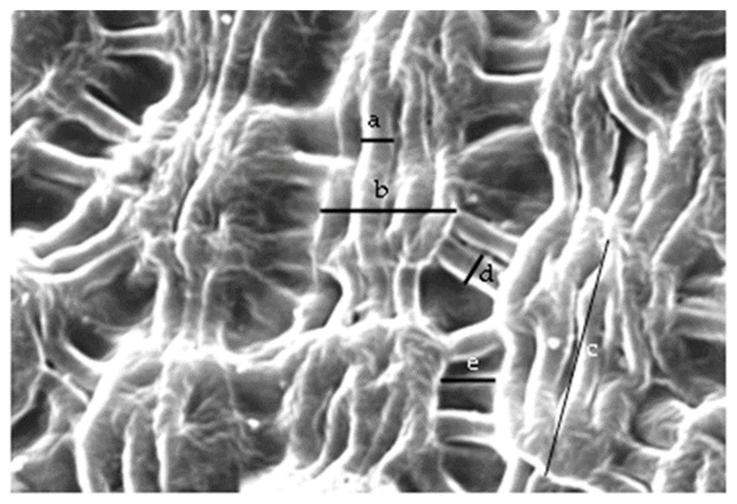
Agriculture | Free Full-Text | Nectar Secretion, Morphology, Anatomy and  Ultrastructure of Floral Nectary in Selected Rubus idaeus L. Varieties |  HTML
Agriculture | Free Full-Text | Nectar Secretion, Morphology, Anatomy and Ultrastructure of Floral Nectary in Selected Rubus idaeus L. Varieties | HTML

INSECT CUTICLE THROUGH THE ELECTRON MICROSCOPE — DISTINGUISHING FACT FROM  ARTIFACT - ScienceDirect
INSECT CUTICLE THROUGH THE ELECTRON MICROSCOPE — DISTINGUISHING FACT FROM ARTIFACT - ScienceDirect

Handbook - The Cuticle

Scanning electron microscopy (SEM) micrographs of the adaxial leaf... |  Download Scientific Diagram
Scanning electron microscopy (SEM) micrographs of the adaxial leaf... | Download Scientific Diagram

INSECT CUTICLE THROUGH THE ELECTRON MICROSCOPE — DISTINGUISHING FACT FROM  ARTIFACT - ScienceDirect
INSECT CUTICLE THROUGH THE ELECTRON MICROSCOPE — DISTINGUISHING FACT FROM ARTIFACT - ScienceDirect

Cuticle and skin cell walls have common and unique roles in grape berry  splitting | Horticulture Research
Cuticle and skin cell walls have common and unique roles in grape berry splitting | Horticulture Research

TEM micrographs of the preecdysial cuticle in Ligia pallasii. (A)... |  Download Scientific Diagram
TEM micrographs of the preecdysial cuticle in Ligia pallasii. (A)... | Download Scientific Diagram

Electron microscopy analysis of the embryonic cuticle of Ore- R (A) and...  | Download Scientific Diagram
Electron microscopy analysis of the embryonic cuticle of Ore- R (A) and... | Download Scientific Diagram

Microscopy of crustacean cuticle: formation of a flexible extracellular  matrix in moulting sea slaters Ligia pallasii | Journal of the Marine  Biological Association of the United Kingdom | Cambridge Core
Microscopy of crustacean cuticle: formation of a flexible extracellular matrix in moulting sea slaters Ligia pallasii | Journal of the Marine Biological Association of the United Kingdom | Cambridge Core

Three‐dimensional imaging of plant cuticle architecture using confocal  scanning laser microscopy - Buda - 2009 - The Plant Journal - Wiley Online  Library
Three‐dimensional imaging of plant cuticle architecture using confocal scanning laser microscopy - Buda - 2009 - The Plant Journal - Wiley Online Library

Secretory cells in active leaf glands, TEM micrographs. 18. Outer... |  Download Scientific Diagram
Secretory cells in active leaf glands, TEM micrographs. 18. Outer... | Download Scientific Diagram

Transmission electron microscopy micrographs of epidermal cells show... |  Download Scientific Diagram
Transmission electron microscopy micrographs of epidermal cells show... | Download Scientific Diagram