Scanning electron microscopy (a–f) and cryomicroscopy (g–h) of isolated... | Download Scientific Diagram

Transmission electron microscopy analysis of leaf cuticle membranes.... | Download Scientific Diagram
![PDF] Ultrastructure of Plant Leaf Cuticles in relation to Sample Preparation as Observed by Transmission Electron Microscopy | Semantic Scholar PDF] Ultrastructure of Plant Leaf Cuticles in relation to Sample Preparation as Observed by Transmission Electron Microscopy | Semantic Scholar](https://d3i71xaburhd42.cloudfront.net/bc063018ecdaa1082ad6f0cc0e3c548fcf9a6323/4-Figure1-1.png)
PDF] Ultrastructure of Plant Leaf Cuticles in relation to Sample Preparation as Observed by Transmission Electron Microscopy | Semantic Scholar
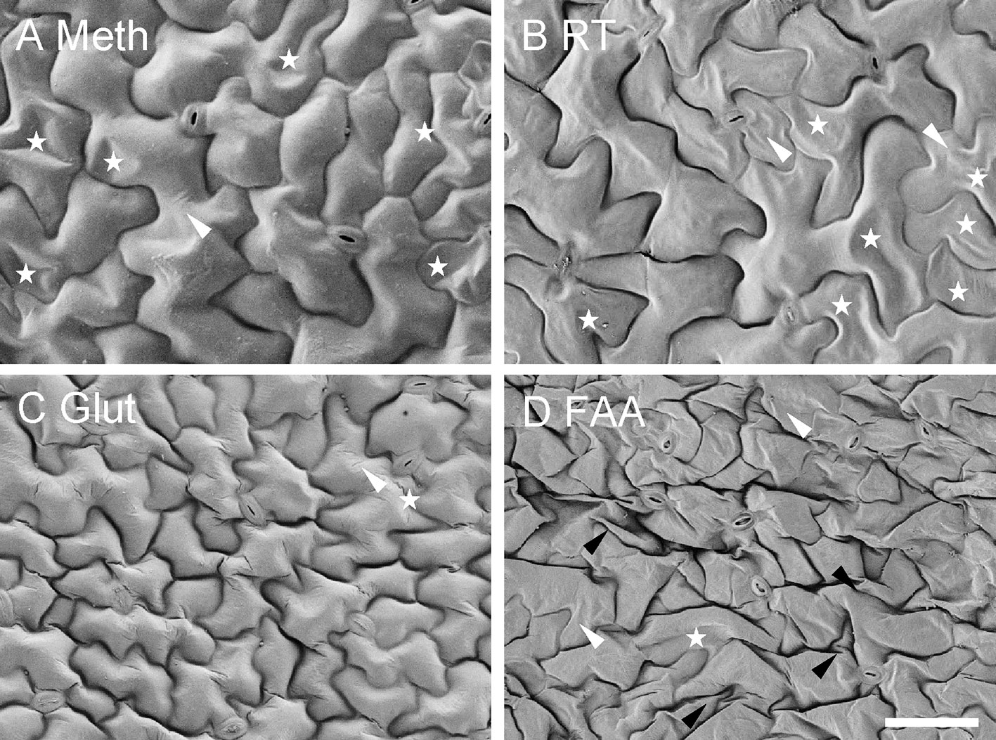
Frontiers | Comparison of Sample Preparation Techniques for Inspection of Leaf Epidermises Using Light Microscopy and Scanning Electronic Microscopy

Transmission electron micrograph of the cuticle surface ofa Panagrellus... | Download Scientific Diagram

Ultrastructure of Plant Leaf Cuticles in relation to Sample Preparation as Observed by Transmission Electron Microscopy
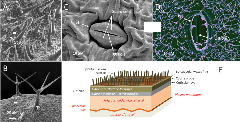
Novel perspectives on stomatal impressions: Rapid and non-invasive surface characterization of plant leaves by scanning electron microscopy | PLOS ONE

TEM analysis of leaf cuticle membranes. The leaf cuticle membrane of a... | Download Scientific Diagram
![PDF] Ultrastructure of Plant Leaf Cuticles in relation to Sample Preparation as Observed by Transmission Electron Microscopy | Semantic Scholar PDF] Ultrastructure of Plant Leaf Cuticles in relation to Sample Preparation as Observed by Transmission Electron Microscopy | Semantic Scholar](https://d3i71xaburhd42.cloudfront.net/bc063018ecdaa1082ad6f0cc0e3c548fcf9a6323/7-Figure3-1.png)
PDF] Ultrastructure of Plant Leaf Cuticles in relation to Sample Preparation as Observed by Transmission Electron Microscopy | Semantic Scholar

Micromorphology and development of the epicuticular structure on the epidermal cell of ginseng leaves - ScienceDirect

Electron microscopy images of dcr mutant epidermal cells. A and B, SEM... | Download Scientific Diagram

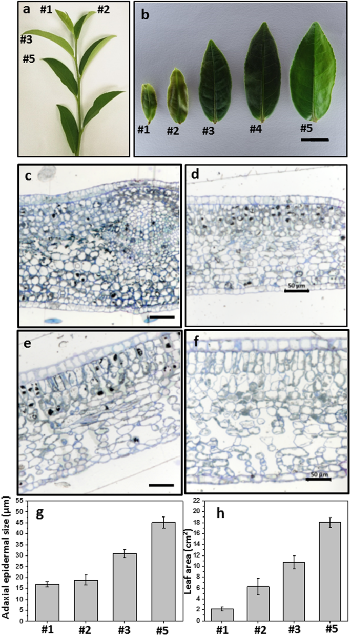
Drought stress modify cuticle of tender tea leaf and mature leaf for transpiration barrier enhancement through common and distinct modes | Scientific Reports

Scanning and transmission electron microscopy of epidermal cell walls... | Download Scientific Diagram
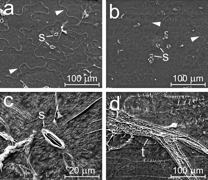
A modified method for enzymatic isolation of and subsequent wax extraction from Arabidopsis thaliana leaf cuticle | Plant Methods | Full Text
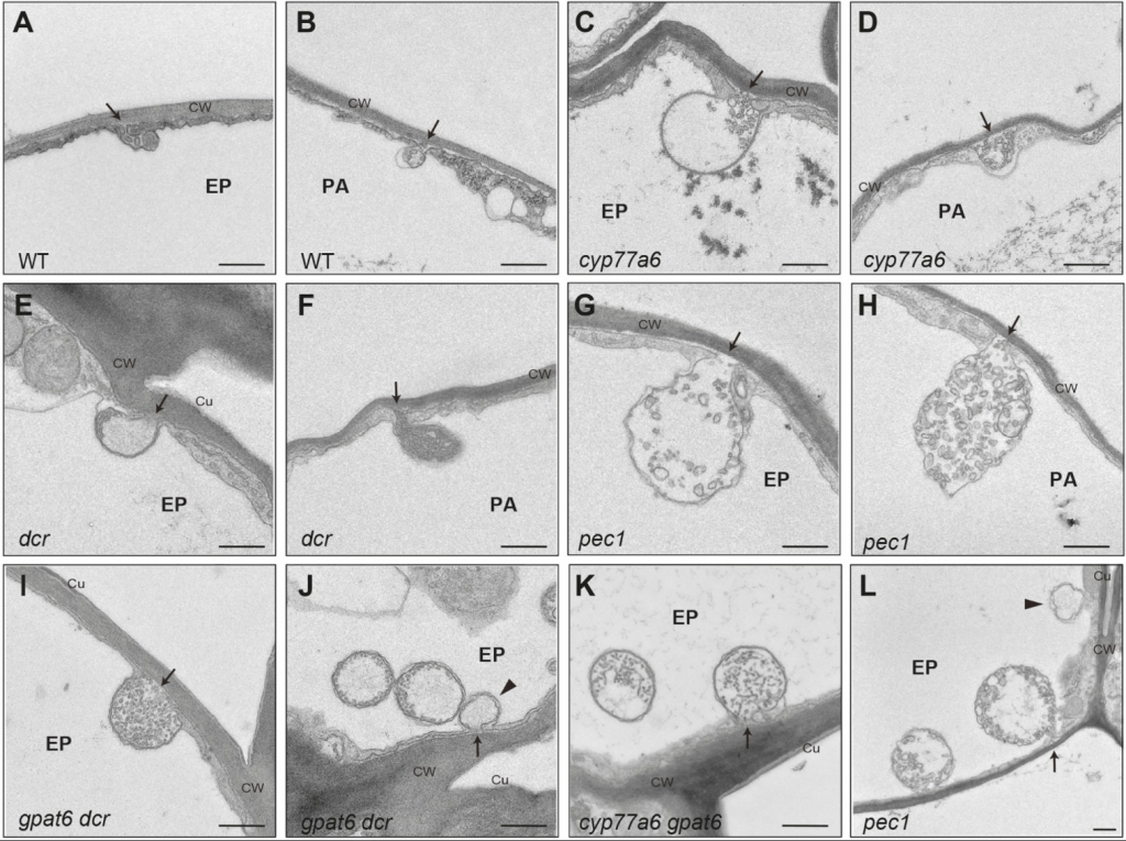
Electron Microscopy Facility University of Lausanne – Page 3 – The goal of the EMF is to promote electron microscopy in Life science. We are part of the Faculty of Biology and

Extracellular lipids of Camelina sativa: Characterization of cutin and suberin reveals typical polyester monomers and novel functionalized dicarboxylic fatty acids | bioRxiv

Transmission electron microscopy micrographs of epidermal cells show... | Download Scientific Diagram
