Home

windswept Disciplinar Purcel clostridium difficile optic microscope Punct căpitan Plicticos

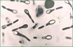
Clostridium difficile (bacteria responsible for hospital acquired  diarrhoea) seen under optical..., Stock Photo, Picture And Rights Managed  Image. Pic. BSI-BSIP-015046-017 | agefotostock
Clostridium difficile (bacteria responsible for hospital acquired diarrhoea) seen under optical..., Stock Photo, Picture And Rights Managed Image. Pic. BSI-BSIP-015046-017 | agefotostock

Real-time tracking of fluorescent magnetic spore–based microrobots for  remote detection of C. diff toxins | Science Advances
Real-time tracking of fluorescent magnetic spore–based microrobots for remote detection of C. diff toxins | Science Advances

Transmission electron microscopy of C. difficile spores. (A) TEM image... |  Download Scientific Diagram
Transmission electron microscopy of C. difficile spores. (A) TEM image... | Download Scientific Diagram

RKI - Consultant Laboratory for Diagnostic Electron Microscopy of  Infectious Pathogens - Clostridium difficile
RKI - Consultant Laboratory for Diagnostic Electron Microscopy of Infectious Pathogens - Clostridium difficile
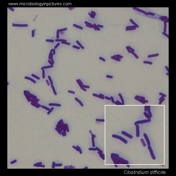
Clostridium difficile microscopy. Clostridium difficile Gram-stain and cell  morphology. Clostridium difficile micrograph, appearance under the  microscope. C.difficile microscopic picture
Clostridium difficile microscopy. Clostridium difficile Gram-stain and cell morphology. Clostridium difficile micrograph, appearance under the microscope. C.difficile microscopic picture

Proteomic and Genomic Characterization of Highly Infectious Clostridium  difficile 630 Spores | Journal of Bacteriology
Proteomic and Genomic Characterization of Highly Infectious Clostridium difficile 630 Spores | Journal of Bacteriology

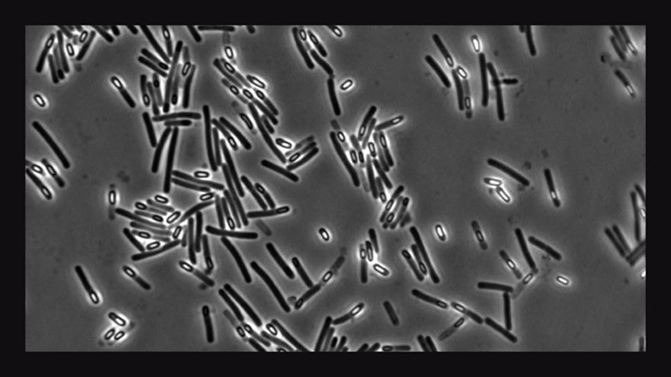
Clostridium difficile. Sporulating culture, Gram-stain and cell morphology.  C.difficile micrograph, appearance under the microscope. Subterminal  spores. C.difficile microscopic picture.
Clostridium difficile. Sporulating culture, Gram-stain and cell morphology. C.difficile micrograph, appearance under the microscope. Subterminal spores. C.difficile microscopic picture.
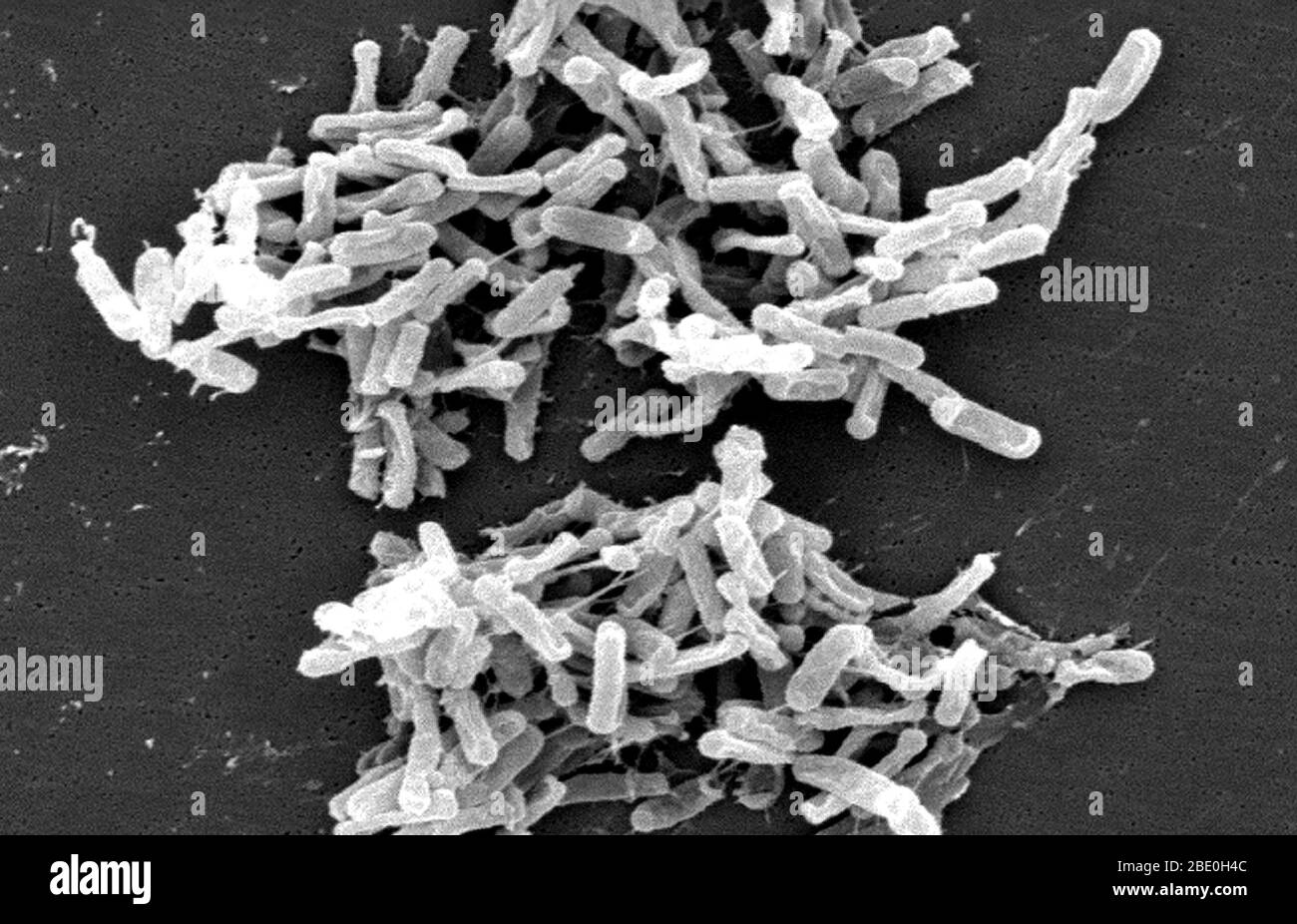
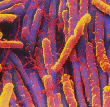
Scanning Electron Micrograph (SEM) showing Gram-positive Clostridium  difficile bacteria. These C. difficile organisms were cultured from a stool  sample obtained during an outbreak of gastrointestinal illness, and  extracted using a .1µm filter.
Scanning Electron Micrograph (SEM) showing Gram-positive Clostridium difficile bacteria. These C. difficile organisms were cultured from a stool sample obtained during an outbreak of gastrointestinal illness, and extracted using a .1µm filter.

Practical observations on the use of fluorescent reporter systems in  Clostridioides difficile | SpringerLink
Practical observations on the use of fluorescent reporter systems in Clostridioides difficile | SpringerLink
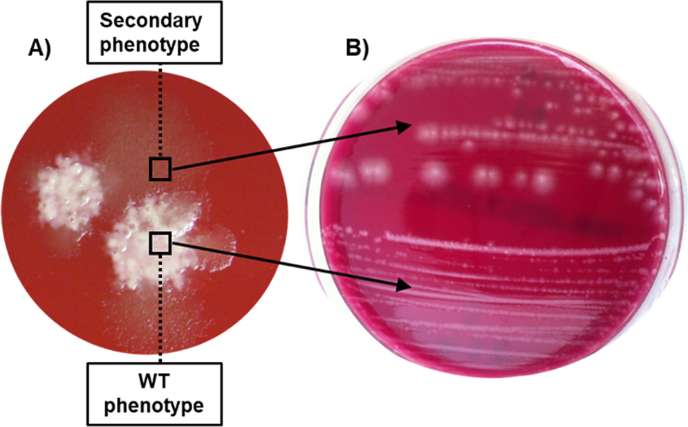
Emergence of a non-sporulating secondary phenotype in Clostridium  (Clostridioides) difficile ribotype 078 isolated from humans and animals |  Scientific Reports
Emergence of a non-sporulating secondary phenotype in Clostridium (Clostridioides) difficile ribotype 078 isolated from humans and animals | Scientific Reports

Effective inhibition of Clostridioides difficile by the novel peptide CM-A  | PLOS ONE
Effective inhibition of Clostridioides difficile by the novel peptide CM-A | PLOS ONE

New UCI-led study uncovers weakness in C. dif | EurekAlert!

Surface Layers of Clostridium difficile Endospores | Journal of Bacteriology

Clostridium difficile (bacteria responsible for hospital acquired  diarrhoea) seen under optical..., Stock Photo, Picture And Rights Managed  Image. Pic. BSI-BSIP-015046-019 | agefotostock
Clostridium difficile (bacteria responsible for hospital acquired diarrhoea) seen under optical..., Stock Photo, Picture And Rights Managed Image. Pic. BSI-BSIP-015046-019 | agefotostock
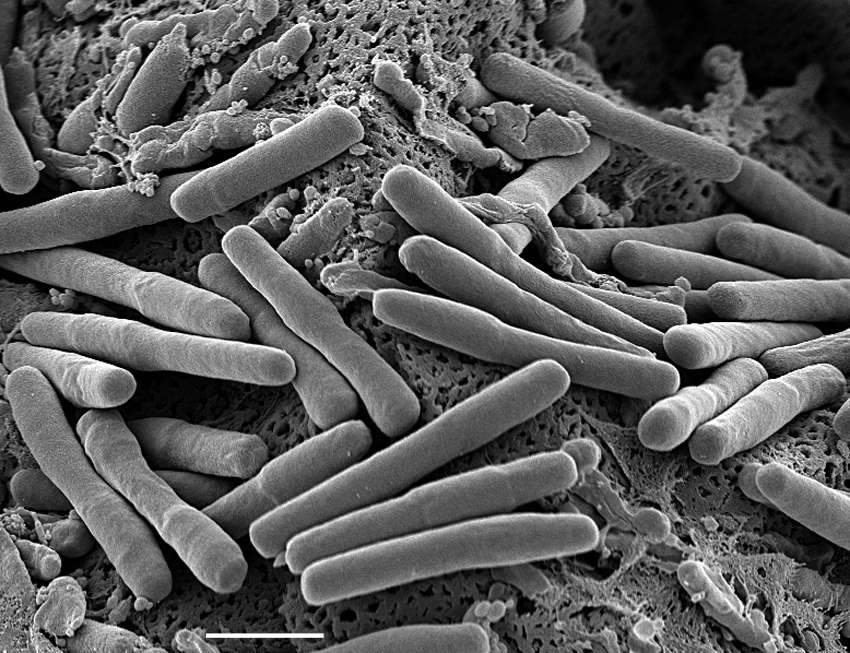
A Revised Understanding of Clostridioides difficile Spore Germination:  Trends in Microbiology
A Revised Understanding of Clostridioides difficile Spore Germination: Trends in Microbiology

Pathology Outlines - Clostridioides difficile

RKI - Consultant Laboratory for Diagnostic Electron Microscopy of  Infectious Pathogens - Clostridium difficile
RKI - Consultant Laboratory for Diagnostic Electron Microscopy of Infectious Pathogens - Clostridium difficile
Clostridium difficile-associated disease - microbewiki

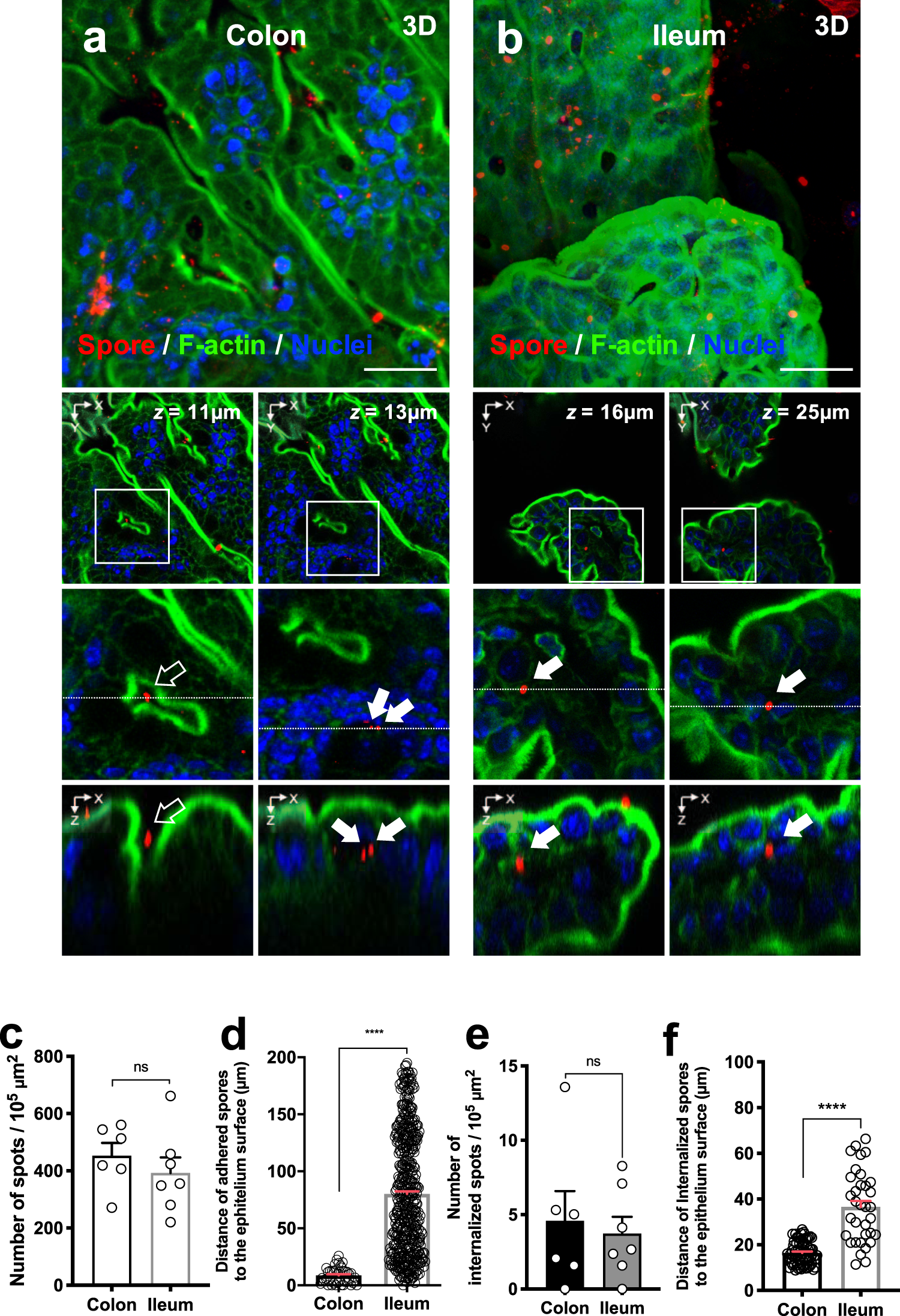
Entry of spores into intestinal epithelial cells contributes to recurrence  of Clostridioides difficile infection | Nature Communications
Entry of spores into intestinal epithelial cells contributes to recurrence of Clostridioides difficile infection | Nature Communications

Pathology Outlines - Clostridioides difficile

IJMS | Free Full-Text | Characterization of an Endolysin Targeting  Clostridioides difficile That Affects Spore Outgrowth | HTML
IJMS | Free Full-Text | Characterization of an Endolysin Targeting Clostridioides difficile That Affects Spore Outgrowth | HTML

Calcium wakes up sleeping C. diff for infection - Futurity

C. difficile was sporulated on SMC solid medium, washed extensively and...  | Download Scientific Diagram
C. difficile was sporulated on SMC solid medium, washed extensively and... | Download Scientific Diagram

Clostridium difficile-associated disease - microbewiki

Butyrate Protects Mice from Clostridium difficile-Induced Colitis through  an HIF-1-Dependent Mechanism - ScienceDirect
Butyrate Protects Mice from Clostridium difficile-Induced Colitis through an HIF-1-Dependent Mechanism - ScienceDirect