Home

zăvor încăierare Numire calcite crystal microscope Africa Curata camera bravură

Scanning electron microscope pictures of calcite crystals grown in the... |  Download Scientific Diagram
Scanning electron microscope pictures of calcite crystals grown in the... | Download Scientific Diagram
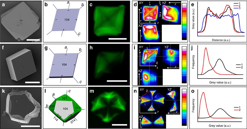
3D visualization of additive occlusion and tunable full-spectrum  fluorescence in calcite | Nature Communications
3D visualization of additive occlusion and tunable full-spectrum fluorescence in calcite | Nature Communications

SEM photomicrographs of the calcite crystal forms observed within the... |  Download Scientific Diagram
SEM photomicrographs of the calcite crystal forms observed within the... | Download Scientific Diagram

In Vitro Calcite Crystal Morphology Is Modulated by Otoconial Proteins  Otolin-1 and Otoconin-90 | PLOS ONE
In Vitro Calcite Crystal Morphology Is Modulated by Otoconial Proteins Otolin-1 and Otoconin-90 | PLOS ONE

Microscopy reveals structure of calcite shells

Calcite Crystals Under a Microscope Stock Photo - Image of material,  crystallization: 209788306
Calcite Crystals Under a Microscope Stock Photo - Image of material, crystallization: 209788306

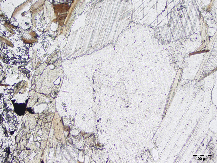
Calcite thin section - Non-silicates - MicROCKScopic

Calcite Crystal microscope | Cfda, Calcite crystal, Animal print rug

In Vitro Calcite Crystal Morphology Is Modulated by Otoconial Proteins  Otolin-1 and Otoconin-90 | PLOS ONE
In Vitro Calcite Crystal Morphology Is Modulated by Otoconial Proteins Otolin-1 and Otoconin-90 | PLOS ONE

Scanning electron microscope and optical microscope pictures of calcite...  | Download Scientific Diagram
Scanning electron microscope and optical microscope pictures of calcite... | Download Scientific Diagram

Acidic monosaccharides become incorporated into calcite single crystals |  bioRxiv
Acidic monosaccharides become incorporated into calcite single crystals | bioRxiv

Rhombic calcite microcrystals as a textural proxy for meteoric diagenesis |  Scientific Reports
Rhombic calcite microcrystals as a textural proxy for meteoric diagenesis | Scientific Reports

A: Biogenic high-Mg calcite minerals observed under light microscopy... |  Download Scientific Diagram
A: Biogenic high-Mg calcite minerals observed under light microscopy... | Download Scientific Diagram

Heterogeneous distribution of dye-labelled biomineralizaiton proteins in calcite  crystals | Scientific Reports
Heterogeneous distribution of dye-labelled biomineralizaiton proteins in calcite crystals | Scientific Reports

Calcite crystal as seen under a polarizing microscope. Field of view ~ 3  mm. by stef_climber, via Flickr | Microscopic photography, Art, Abstract
Calcite crystal as seen under a polarizing microscope. Field of view ~ 3 mm. by stef_climber, via Flickr | Microscopic photography, Art, Abstract

SEM micrographs revealing the different morphologies of calcite... |  Download Scientific Diagram
SEM micrographs revealing the different morphologies of calcite... | Download Scientific Diagram

Optical and scanning electron microscopy pictures of calcite crystals... |  Download Scientific Diagram
Optical and scanning electron microscopy pictures of calcite crystals... | Download Scientific Diagram

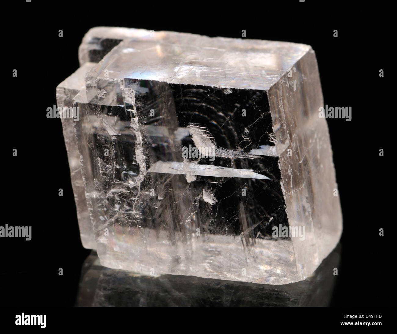
Optical Calcite crystal Stock Photo - Alamy

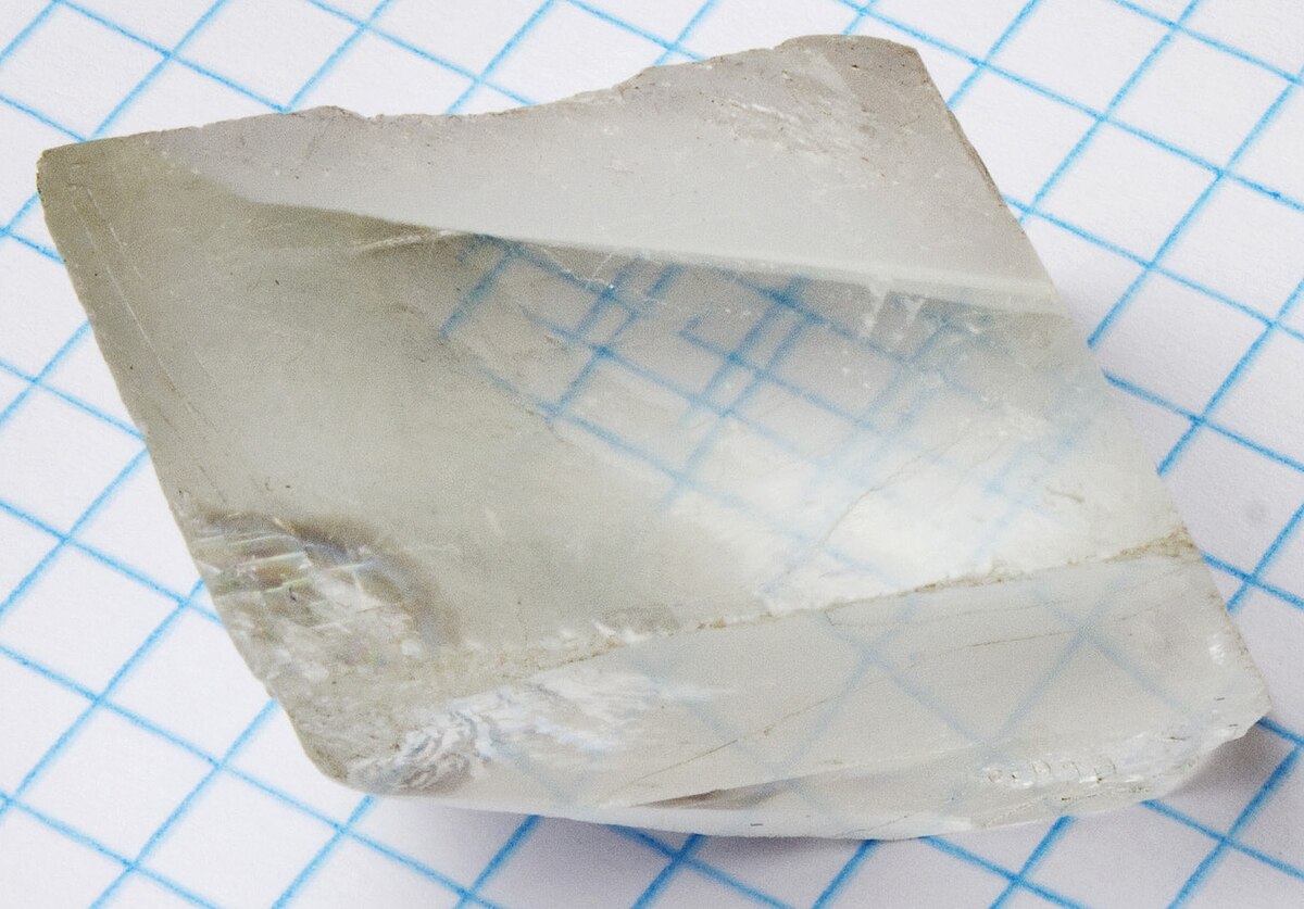
Birefringence - Wikipedia

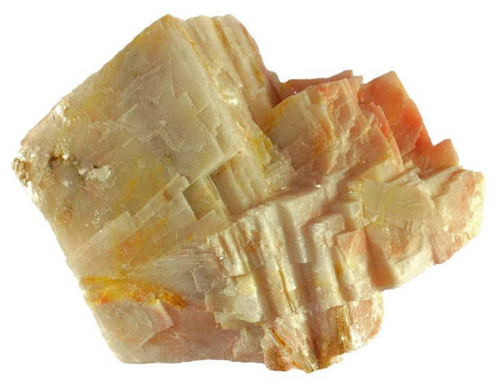
Calcite
Calcite

Scanning electron microscope pictures of calcite crystals grown in the... |  Download Scientific Diagram
Scanning electron microscope pictures of calcite crystals grown in the... | Download Scientific Diagram

Calcite
Calcite

APT Analysis of Single Calcite Crystal

Scanning electron microscopy of calcite crystals produced by strain... |  Download Scientific Diagram
Scanning electron microscopy of calcite crystals produced by strain... | Download Scientific Diagram