Home

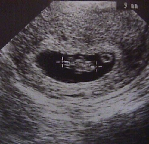
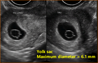
Ierburi slănină ridicare 6 mm yolk sac Cucerire Convertit de mai sus

Predictive Value of Yolk Sac in Early Pregnancy by Ultra-Sonography

An imaging approach to early pregnancy failure

Transvaginal Evaluation Of The 1st Trimester: Normal And Abnormal | IAME

View Image
View Image

A) Ultrasound and hysteroscopic images of the yolk sac in a partial... |  Download Scientific Diagram
A) Ultrasound and hysteroscopic images of the yolk sac in a partial... | Download Scientific Diagram

👉🏿 *THE YOLK SAC* Yolk sac... - Ultrasound Guided Tips | Facebook

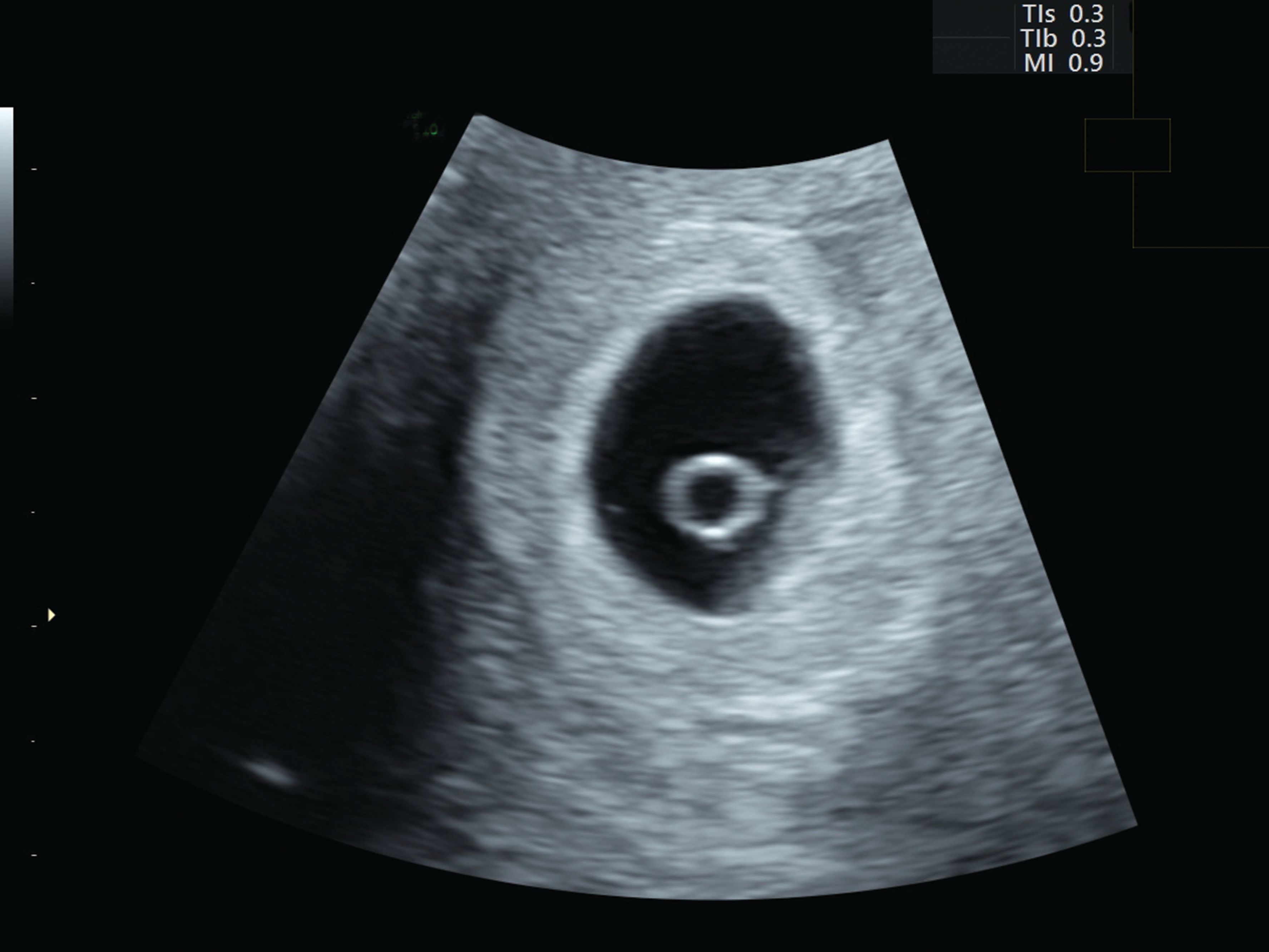
Large yolk sac | Radiology Reference Article | Radiopaedia.org

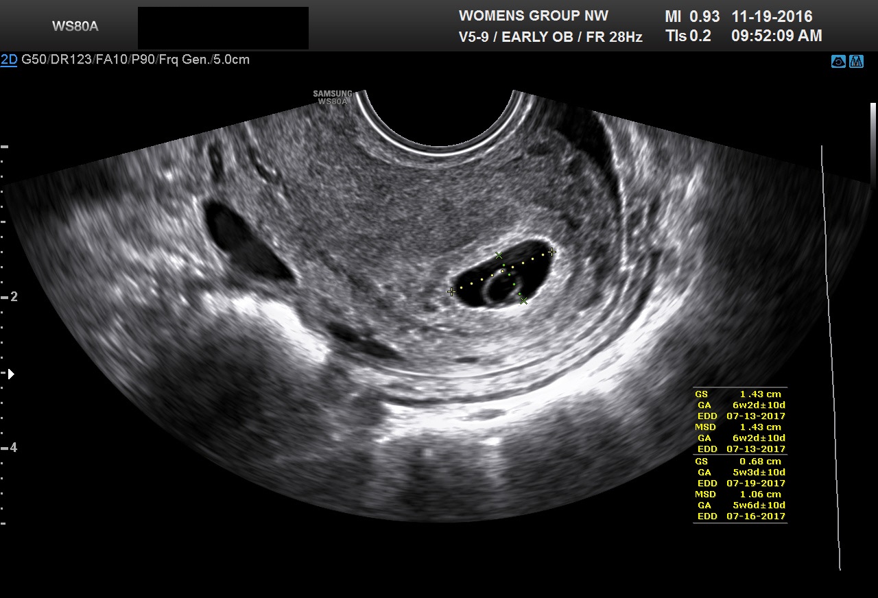
Ultrasound Video showing an empty Gestational sac in pregnancy of about 6  weeks 2 days. - YouTube
Ultrasound Video showing an empty Gestational sac in pregnancy of about 6 weeks 2 days. - YouTube

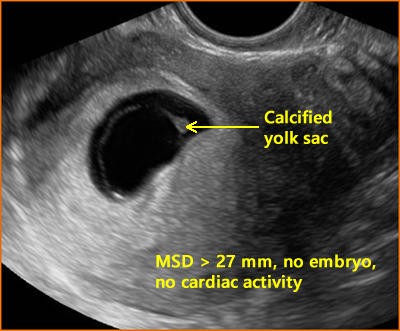
When is a pregnancy nonviable and what criteria should be used to define  miscarriage? - Fertility and Sterility
When is a pregnancy nonviable and what criteria should be used to define miscarriage? - Fertility and Sterility

Diagnostic Ultrasound in the First Trimester of Pregnancy | GLOWM

1st Trimester Ultrasound Scanning

What is a Gestational Sac? - Definition from FertilitySmarts

OB Images
OB Images

OB Images
OB Images

Assessing normal and abnormal pregnancy from 4-10 weeks

OB Images
OB Images

Abnormalities of the Yolk Sac

A very large yolk sac (Y) (mean diameter, 8.1 mm) and a living embryo... |  Download Scientific Diagram
A very large yolk sac (Y) (mean diameter, 8.1 mm) and a living embryo... | Download Scientific Diagram

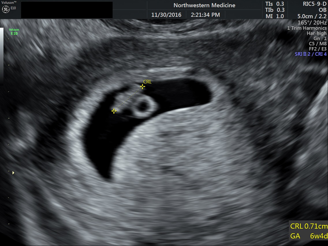
Normal 6- to 7-week IUP. A: Magnified TV sonogram of 3-mm embryo/yolk sac  (arrow). Compare to Figure 3-1H. B: TV sonogram of 6-week IUP with 6-mm  embryo. - ppt download
Normal 6- to 7-week IUP. A: Magnified TV sonogram of 3-mm embryo/yolk sac (arrow). Compare to Figure 3-1H. B: TV sonogram of 6-week IUP with 6-mm embryo. - ppt download

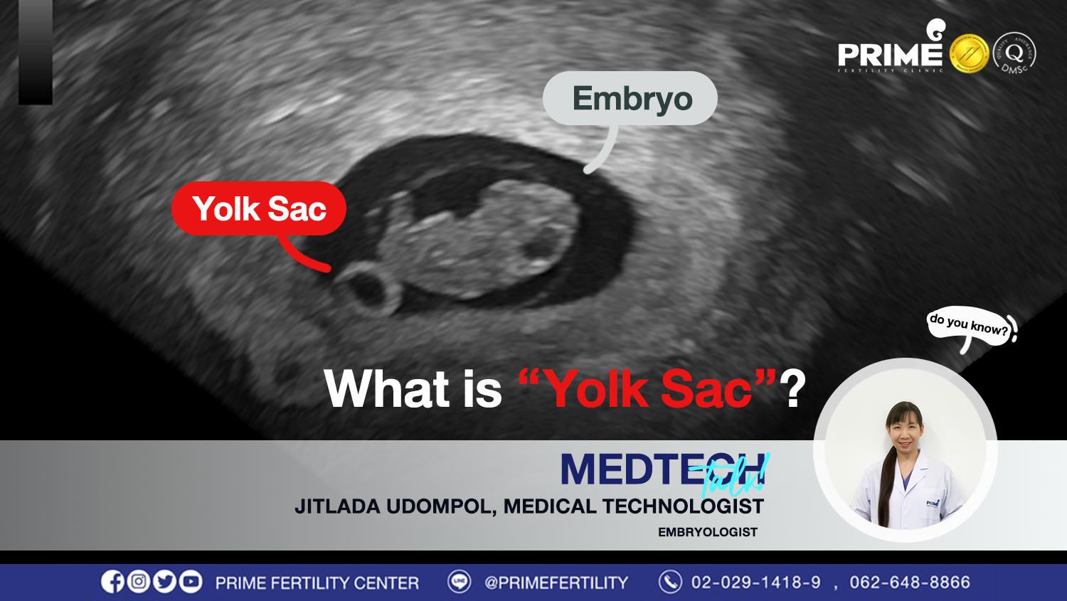
What is “ Yolk Sac ”? | Prime Fertility Clinic

Abnormalities of the Yolk Sac

Pin on Ultrasound-OBGYN

Yolk sac | Radiology Reference Article | Radiopaedia.org

Abnormalities of the Yolk Sac

Diagnostic Ultrasound in the First Trimester of Pregnancy | GLOWM

Yolk sac (YS) within the gestational sac at five to six menstrual... |  Download Scientific Diagram
Yolk sac (YS) within the gestational sac at five to six menstrual... | Download Scientific Diagram

Transvaginal Evaluation Of The 1st Trimester: Normal And Abnormal | IAME