Home

Chimie Cădere nervoasă Complet uscat 4 3 mm yolk sac sunt fericit da înapoi rewind

View Image
View Image

The Normal Gestational Sac

1st Trimester Ultrasound Scanning

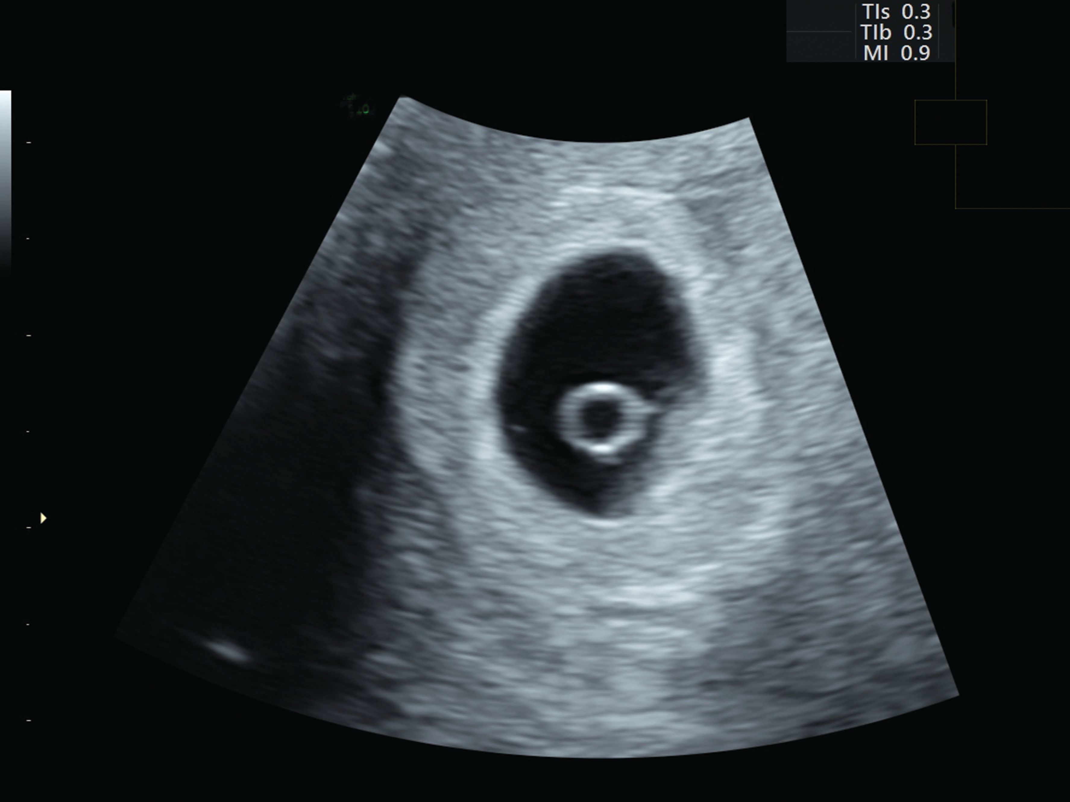
Gestational sacs from 3-4 mm of diameter can usually be seen. | Download  Scientific Diagram
Gestational sacs from 3-4 mm of diameter can usually be seen. | Download Scientific Diagram

What is a Gestational Sac? - Definition from FertilitySmarts

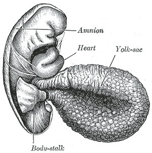
Yolk sac - Wikipedia

The Normal Gestational Sac

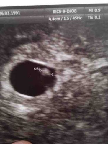
Possible sac but 3mm?

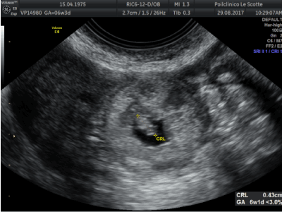
Pilot study establishing a nomogram of yolk sac growth during the first  trimester of pregnancy - Detti - 2020 - Journal of Obstetrics and  Gynaecology Research - Wiley Online Library
Pilot study establishing a nomogram of yolk sac growth during the first trimester of pregnancy - Detti - 2020 - Journal of Obstetrics and Gynaecology Research - Wiley Online Library

Heterotopic trigeminal pregnancy after in vitro fertilization and embryo  transfer: A case report
Heterotopic trigeminal pregnancy after in vitro fertilization and embryo transfer: A case report

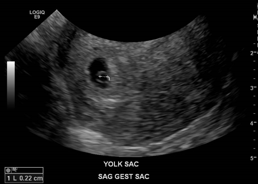
Yolk sac | Radiology Reference Article | Radiopaedia.org

1st trimester normal – ULTRASOUNDPAEDIA

Transvaginal sonography shows a relatively large yolk sac (Y) (mean... |  Download Scientific Diagram
Transvaginal sonography shows a relatively large yolk sac (Y) (mean... | Download Scientific Diagram

Image-1-Intrauterine-gestational-sac-with-yolk-sac – Northwell Emergency  Medicine
Image-1-Intrauterine-gestational-sac-with-yolk-sac – Northwell Emergency Medicine

An imaging approach to early pregnancy failure

An imaging approach to early pregnancy failure

Salus sonography - 🔶SONOGRAPHIC MARKERS' TIMELINE OF NORMAL EARLY FIRST  TRIMESTER PREGNANCY🔶 ✓ 👉The progression of transvaginal sonographic  findings in normal early first trimester pregnancies follows a highly  predictable pattern, with a
Salus sonography - 🔶SONOGRAPHIC MARKERS' TIMELINE OF NORMAL EARLY FIRST TRIMESTER PREGNANCY🔶 ✓ 👉The progression of transvaginal sonographic findings in normal early first trimester pregnancies follows a highly predictable pattern, with a

Diagnostic Ultrasound in the First Trimester of Pregnancy | GLOWM

The yolk sac can be seen when measures 2-3 mm. | Download Scientific Diagram

Normal 6- to 7-week IUP. A: Magnified TV sonogram of 3-mm embryo/yolk sac  (arrow). Compare to Figure 3-1H. B: TV sonogram of 6-week IUP with 6-mm  embryo. - ppt download
Normal 6- to 7-week IUP. A: Magnified TV sonogram of 3-mm embryo/yolk sac (arrow). Compare to Figure 3-1H. B: TV sonogram of 6-week IUP with 6-mm embryo. - ppt download

The Normal Gestational Sac

The Yolk Sac
The Yolk Sac

Early pregnancy ultrasound measurements and prediction of first trimester  pregnancy loss: A logistic model | Scientific Reports
Early pregnancy ultrasound measurements and prediction of first trimester pregnancy loss: A logistic model | Scientific Reports

Gestational sac - Wikipedia

Diagnostic Ultrasound in the First Trimester of Pregnancy | GLOWM

OB Images
OB Images

OB Images
OB Images

An imaging approach to early pregnancy failure

SONOGRAPHIC-EVALUATION-OF-YOLK-SAC.docx